
-
Product Name
Anti-E Cadherin (7H12) Mouse antibody
- Documents
-
Description
E Cadherin (7H12) Mouse monoclonal antibody
-
Tested applications
WB, IHC-P, FC
-
Species reactivity
Human, Mouse, Monkey
-
Isotype
Mouse IgG1
-
Preparation
Antigen: Purified recombinant fragment of human CDH1 expressed in E. Coli.
-
Clonality
Monoclonal
-
Formulation
Ascitic fluid containing 0.03% sodium azide.
-
Storage instructions
Store at 4°C short term. Store at -20°C long term. Avoid freeze / thaw cycle.
-
Applications
WB: 1/500 - 1/2000
IHC: 1/200 - 1/1000
FC: 1/200 - 1/400
ELISA: 1/10000
-
Validations
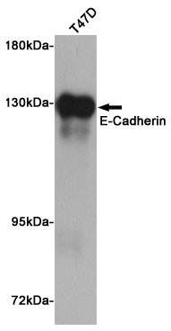
Western blot analysis of extract from T47D cells using E-Cadherin Mouse mAb at 1:1000 dilution. Predicted band size: 135KDa. Observed band size: 135KDa.
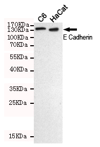
Western blot detection of E-Cadherin in C6 and HaCat cell lysates using E Cadherin mouse mAb (dilution 1:500).Predicted band size:135KDa.Observed band size:135KDa.
-
Background
Swiss-Prot Acc.P12830.E-Cadherin is a 120 kDa transmembrane glycoprotein that is localized in the adherens junctions of epithelial cells. There, it interacts with the cytoskeleton through the associated cytoplasmic catenin proteins. In addition to being a calcium-dependent adhesion molecule, E-Cadherin is also a critical regulator of epithelial junction formation. Its association with catenins is necessary for cell-cell adhesion. These E-cadherin/catenin complexes associate with corical actin bundles at both the zonula adherens and the lateral adhesion plaques. Tyrosine phosphorylation can disrupt these complexes, leading to changes in cell adhesion properties. E-Cadherin expression is often down-regulated in highly invasive, poorly differentiated carcinomas. Increased expression of E-Cadherin in these cells reduces invasiveness. Thus, loss of expression or function of E-Cadherin appears to be an important step in tumorigenic progression.Tissue specificity: Non-neural epithelial tissues.
Related Products / Services
Please note: All products are "FOR RESEARCH USE ONLY AND ARE NOT INTENDED FOR DIAGNOSTIC OR THERAPEUTIC USE"