
-
Product Name
E-cadherin antibody
- Documents
-
Description
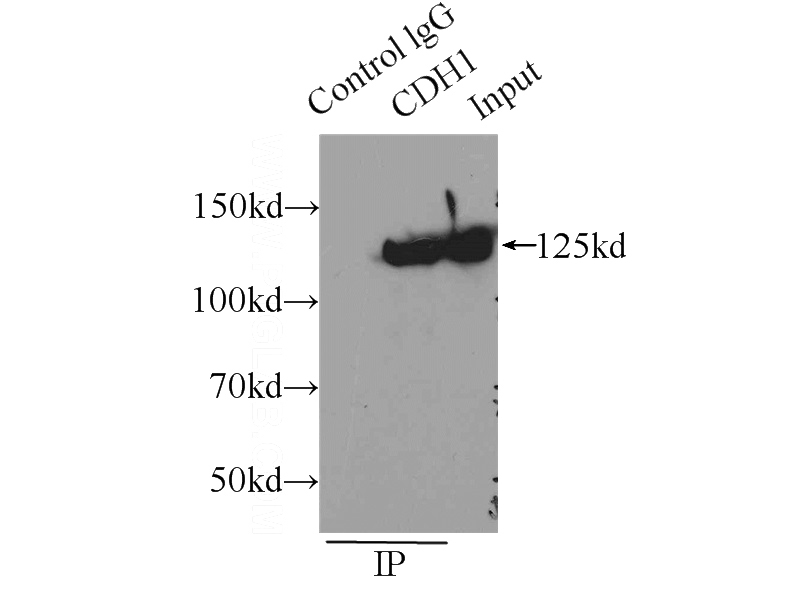
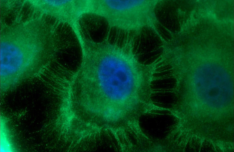
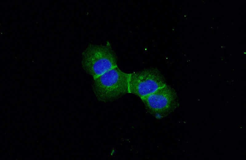
E-cadherin Rabbit Polyclonal antibody. Positive IP detected in A431 cells. Positive WB detected in mouse testis tissue, A431 cells, DU 145 cells, MCF7 cells, PC-3 cells. Positive FC detected in HepG2 cells. Positive IHC detected in human skin tissue, human breast cancer tissue, human colon tissue, human hepat℃irrhosis tissue, human liver cancer tissue, mouse liver tissue. Positive IF detected in A431 cells. Observed molecular weight by Western-blot: 125 kDa
-
Tested applications
ELISA, IHC, WB, IP, FC, IF
-
Species reactivity
Human,Mouse,Rat; other species not tested.
-
Alternative names
Arc 1 antibody; Cadherin 1 antibody; CD324 antibody; CDH1 antibody; CDHE antibody; E cadherin antibody; ECAD antibody; E-cadherin antibody; LCAM antibody; UVO antibody; Uvomorulin antibody
-
Isotype
Rabbit IgG
-
Preparation
This antibody was obtained by immunization of E-cadherin recombinant protein (Accession Number: BC141838). Purification method: Antigen affinity purified.
-
Clonality
Polyclonal
-
Formulation
PBS with 0.02% sodium azide and 50% glycerol pH 7.3.
-
Storage instructions
Store at -20℃. DO NOT ALIQUOT
-
Applications
Recommended Dilution:
WB: 1:500-1:5000
IP: 1:500-1:5000
IHC: 1:20-1:200
IF: 1:20-1:200
-
Validations
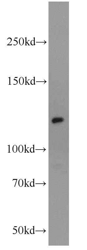
mouse testis tissue were subjected to SDS PAGE followed by western blot with Catalog No:110288(E-cadherin antibody) at dilution of 1:1000
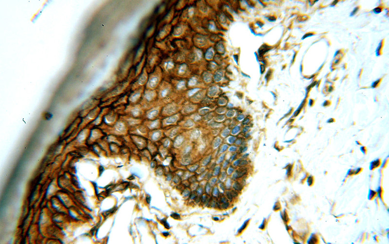
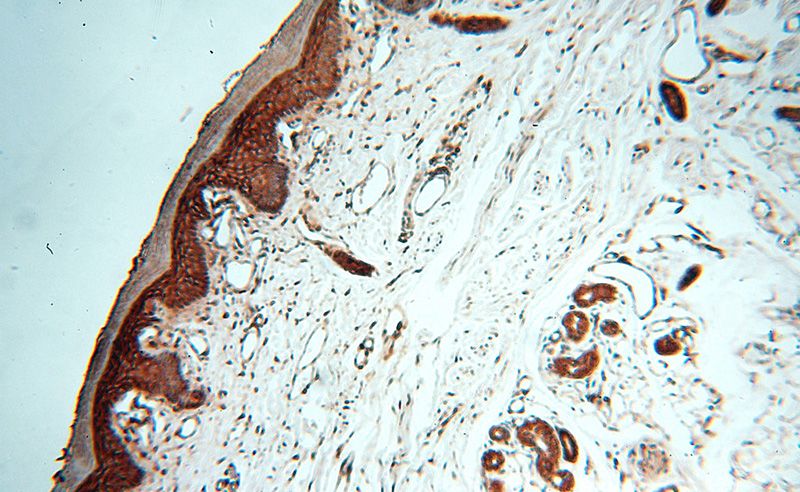
Immunohistochemical of paraffin-embedded human skin using Catalog No:110288(E-cadherin antibody) at dilution of 1:50 (under 40x lens)
Immunohistochemical of paraffin-embedded human skin using Catalog No:110288(E-cadherin antibody) at dilution of 1:50 (under 10x lens)
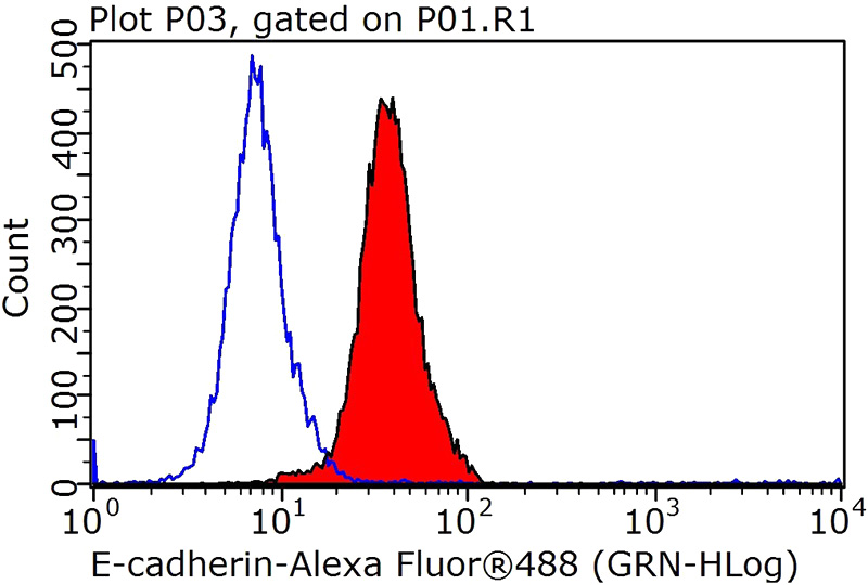
1X10^6 HepG2 cells were stained with .2ug E-cadherin antibody (Catalog No:110288, red) and control antibody (blue). Fixed with 90% MeOH blocked with 3% BSA (30 min). Alexa Fluor 488-congugated AffiniPure Goat Anti-Rabbit IgG(H+L) with dilution 1:1000.
IP Result of anti-E-cadherin (IP:Catalog No:110288, 3ug; Detection:Catalog No:110288 1:1000) with A431 cells lysate 3000ug.
Immunofluorescent analysis of A431 cells using Catalog No:110288(E-cadherin Antibody) at dilution of 1:25 and Alexa Fluor 488-congugated AffiniPure Goat Anti-Rabbit IgG(H+L)
Immunofluorescent analysis of (-20°C Ethanol) fixed A431 cells using Catalog No:110288(E-cadherin Antibody) at dilution of 1:50 and Alexa Fluor 488-congugated AffiniPure Goat Anti-Rabbit IgG(H+L)
-
Background
Cadherins are a family of transmembrane glycoproteins that mediate calcium-dependent cell-cell adhesion and play an important role in the maintenance of normal tissue architecture. E-cadherin (epithelial cadherin), also known as CDH1 (cadherin 1) or CAM 120/80, is a classical member of the cadherin superfamily which also include N-, P-, R-, and B-cadherins. It has been regarded as a marker for spermatogonial stem cells in mice(PMID:23509752). E-cadherin is expressed on the cell surface in most epithelial tissues. The extracellular region of E-cadherin establishes calcium-dependent homophilic trans binding, providing specific interaction with adjacent cells, while the cytoplasmic domain is connected to the actin cytoskeleton through the interaction with p120-, α-, β-, and γ-catenin (plakoglobin). E-cadherin is important in the maintenance of the epithelial integrity, and is involved in mechanisms regulating proliferation, differentiation, and survival of epithelial cell. E-cadherin may also play a role in tumorigenesis. It is considered to be an invasion suppressor protein and its loss is an indicator of high tumor aggressiveness. E-cadherin is sensitive to trypsin digestion in the absence of Ca2+. This polyclonal antibody recognizes 120-kDa intact E-cadherin and its 80-kDa trypsin-cleaved fragment.
-
References
- Zhu L, Qin H, Li PY. Response gene to complement-32 enhances metastatic phenotype by mediating transforming growth factor beta-induced epithelial-mesenchymal transition in human pancreatic cancer cell line BxPC-3. Journal of experimental & clinical cancer research : CR. 31:29. 2012.
- Chen XY, Gu XT, Saiyin H. Brain-selective kinase 2 (BRSK2) phosphorylation on PCTAIRE1 negatively regulates glucose-stimulated insulin secretion in pancreatic β-cells. The Journal of biological chemistry. 287(36):30368-75. 2012.
- Ding J, Jin W, Chen C, Shao Z, Wu J. Tumor associated macrophage × cancer cell hybrids may acquire cancer stem cell properties in breast cancer. PloS one. 7(7):e41942. 2012.
- Song Y, Xue L, Du S. Caveolin-1 knockdown is associated with the metastasis and proliferation of human lung cancer cell line NCI-H460. Biomedicine & pharmacotherapy = Biomédecine & pharmacothérapie. 66(6):439-47. 2012.
- Liu C, Zhang A, Guo J. Identification of human host proteins contributing to H5N1 influenza virus propagation by membrane proteomics. Journal of proteome research. 11(11):5396-405. 2012.
- Zha L, Zhang J, Tang W. HMGA2 elicits EMT by activating the Wnt/β-catenin pathway in gastric cancer. Digestive diseases and sciences. 58(3):724-33. 2013.
- Ning Q, Liu C, Hou L. Vascular endothelial growth factor receptor-1 activation promotes migration and invasion of breast cancer cells through epithelial-mesenchymal transition. PloS one. 8(6):e65217. 2013.
- Liu N, Li Y, Su S, Wang N, Wang H, Li J. Inhibition of cell migration by ouabain in the A549 human lung cancer cell line. Oncology letters. 6(2):475-479. 2013.
Related Products / Services
Please note: All products are "FOR RESEARCH USE ONLY AND ARE NOT INTENDED FOR DIAGNOSTIC OR THERAPEUTIC USE"