
-
Product Name
Anti-EGFR (6H11) Mouse antibody
- Documents
-
Description
EGFR (6H11) Mouse monoclonal antibody
-
Tested applications
WB, ICC/IF, IP
-
Species reactivity
Human, Monkey
-
Alternative names
Avian erythroblastic leukemia viral (v erb b) oncogene homolog;Cell growth inhibiting protein 40;Cell proliferation inducing protein 61;EGF R;EGFR;EGFR_HUMAN;Epidermal growth factor receptor (avian erythroblastic leukemia viral (v erb b) oncogene homolog) antibody
-
Isotype
Mouse IgG1
-
Preparation
Antigen: Purified recombinant human EGFR protein fragments expressed in E.coli.
-
Clonality
Monoclonal
-
Formulation
Purified mouse monoclonal antibody in PBS(pH 7.4) containing with 0.02% sodium azide 0.1%BSA and 50% glycerol.
-
Storage instructions
Store at 4°C short term. Store at -20°C long term. Avoid freeze / thaw cycle.
-
Applications
WB: 1/2000
;ICC: 1/200
-
Validations
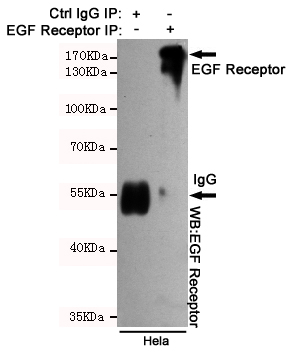
Immunoprecipitation analysis of Hela cell lysates using EGFR mouse mAb.
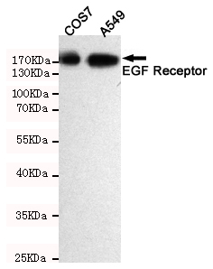
Western blot detection of EGFR in A549 and COS7 cell lysates using EGFR mouse mAb(dilution 1:2000).Predicted band size:134 Kda.Observed band size:175KDa.
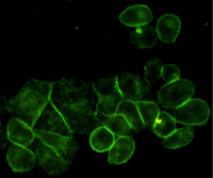
Immunocytochemistry staining of HeLa cells using EGFR mouse mAb (dilution 1:200).
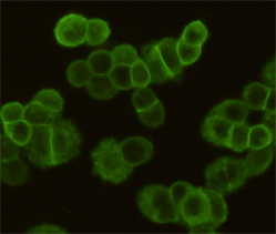
Immunocytochemistry staining of MDA-MB-468 cells fixed with 4% Paraformaldehyde and using EGFR mouse mAb (dilution 1:200).
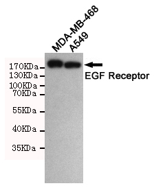
Western blot detection of EGFR in A549 and MDA-MB-468 cell lysates using EGFR mouse mAb(dilution 1:1000).Predicted band size:134 Kda.Observed band size:175KDa.
-
Background
Swiss-Prot Acc.P00533.EGFR is a receptor tyrosine kinase. Receptor for epidermal growth factor (EGF) and related growth factors including TGF-alpha, amphiregulin, betacellulin, heparin-binding EGF-like growth factor, GP30 and vaccinia virus growth factor. Is involved in the control of cell growth and differentiation. . A single-pass transmembrane tyrosine kinase. Ligand binding to this receptor results in receptor dimerization, autophosphorylation (in trans), activation of various downstream signaling molecules and lysosomal degradation.
Related Products / Services
Please note: All products are "FOR RESEARCH USE ONLY AND ARE NOT INTENDED FOR DIAGNOSTIC OR THERAPEUTIC USE"