-
Product Name
UCHL1/PGP9.5 antibody
- Documents
-
Description
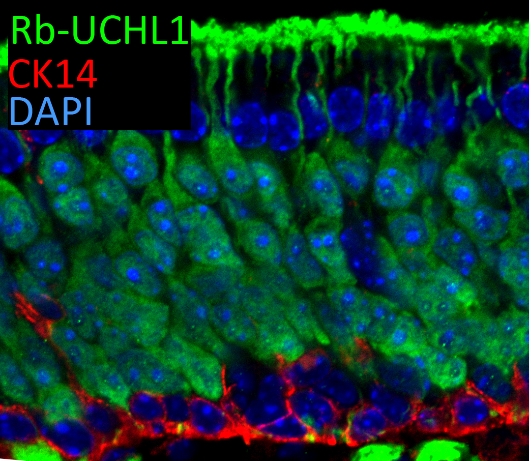
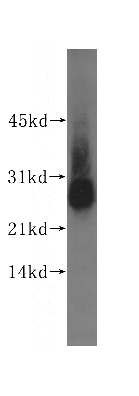
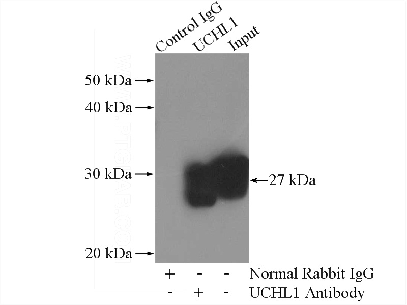
UCHL1/PGP9.5 Rabbit Polyclonal antibody. Positive WB detected in A549 cells, rat lung tissue, SH-SY5Y cells. Positive IP detected in mouse brain tissue. Positive IF detected in mouse olfactory epithelium tissue. Observed molecular weight by Western-blot: 27 kDa
-
Tested applications
ELISA, WB, IP, IF
-
Species reactivity
Human,Mouse,Rat; other species not tested.
-
Alternative names
Neuron cytoplasmic protein 9.5 antibody; PARK5 antibody; PGP 9.5 antibody; PGP9.5 antibody; Ubiquitin thioesterase L1 antibody; Uch L1 antibody; UCHL1 antibody
-
Isotype
Rabbit IgG
-
Preparation
This antibody was obtained by immunization of UCHL1/PGP9.5 recombinant protein (Accession Number: NM_004181). Purification method: Antigen affinity purified.
-
Clonality
Polyclonal
-
Formulation
PBS with 0.02% sodium azide and 50% glycerol pH 7.3.
-
Storage instructions
Store at -20℃. DO NOT ALIQUOT
-
Applications
Recommended Dilution:
WB: 1:500-1:5000
IP: 1:1000-1:10000
IF: N/A
-
Validations

IF result of UCHL1 (Catalog No:116671, 1:300) with 1% PLP fixed adult mouse olfactory epithelium. (Red: CK14; Green: UCHL1; Blue: DAPI). By Brian Lin, Tufts University.

A549 cells were subjected to SDS PAGE followed by western blot with Catalog No:116671(UCHL1 antibody) at dilution of 1:400

IP Result of anti-UCHL1 (IP:Catalog No:116671, 4ug; Detection:Catalog No:116671 1:2000) with mouse brain tissue lysate 4000ug.
-
Background
UCHL1(Ubiquitin carboxyl-terminal hydrolase isozyme L1) is a member of a gene family whose products hydrolyze small C-terminal adducts of ubiquitin to generate the ubiquitin monomer. Expression of UCHL1 is highly specific to neurons and to cells of the diffuse neuroendocrine system and their tumors. It is present in all neurons(PMID: 6343558). This protein is present in brain at concentrations at least 50 times greater than in other organs and is a major protein component of neuronal cytoplasm (PMID:7217993). And UCHL1 is a Parkinson's disease susceptibility gene (PMID:15048890).
-
References
- Jara JH, Genç B, Cox GA. Corticospinal Motor Neurons Are Susceptible to Increased ER Stress and Display Profound Degeneration in the Absence of UCHL1 Function. Cerebral cortex (New York, N.Y. : 1991). 25(11):4259-72. 2015.
- Genç B, Lagrimas AK, Kuru P. Visualization of Sensory Neurons and Their Projections in an Upper Motor Neuron Reporter Line. PloS one. 10(7):e0132815. 2015.
- Huang SH, Wang L, Chi F. Circulating brain microvascular endothelial cells (cBMECs) as potential biomarkers of the blood-brain barrier disorders caused by microbial and non-microbial factors. PloS one. 8(4):e62164. 2013.
Related Products / Services
Please note: All products are "FOR RESEARCH USE ONLY AND ARE NOT INTENDED FOR DIAGNOSTIC OR THERAPEUTIC USE"
