
-
Product Name
IGF2BP3 antibody
- Documents
-
Description
IGF2BP3 Rabbit Polyclonal antibody. Positive IF detected in MCF-7 cells. Positive IHC detected in human pancreas cancer tissue. Positive WB detected in HepG2 cells, BxPC-3 cells, HeLa cells, mouse brain tissue. Positive IP detected in HeLa cells. Observed molecular weight by Western-blot: 64 kDa
-
Tested applications
ELISA, WB, IHC, IF, IP
-
Species reactivity
Human, Mouse; other species not tested.
-
Alternative names
DKFZp686F1078 antibody; hKOC antibody; IGF II mRNA binding protein 3 antibody; IGF2 mRNA binding protein 3 antibody; IGF2BP3 antibody; IMP 3 antibody; IMP3 antibody; KOC1 antibody; VICKZ family member 3 antibody; VICKZ3 antibody
- Immunogen
-
Isotype
Rabbit IgG
-
Preparation
This antibody was obtained by immunization of IGF2BP3 recombinant protein (Accession Number: NM_006547). Purification method: Antigen affinity purified.
-
Clonality
Polyclonal
-
Formulation
PBS with 0.02% sodium azide and 50% glycerol pH 7.3.
-
Storage instructions
Store at -20℃. DO NOT ALIQUOT
-
Applications
Recommended Dilution:
WB: 1:500-1:5000
IP: 1:500-1:5000
IHC: 1:20-1:200
IF: 1:10-1:100
-
Validations
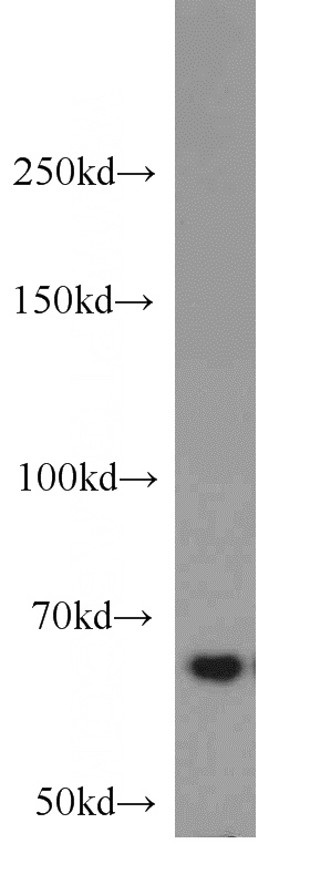
HepG2 cells were subjected to SDS PAGE followed by western blot with Catalog No:111681(IGF2BP3 antibody) at dilution of 1:1000
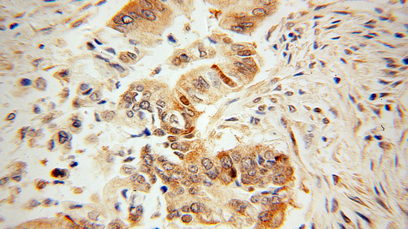
Immunohistochemical of paraffin-embedded human pancreas cancer using Catalog No:111681(IGF2BP3 antibody) at dilution of 1:100 (under 40x lens)
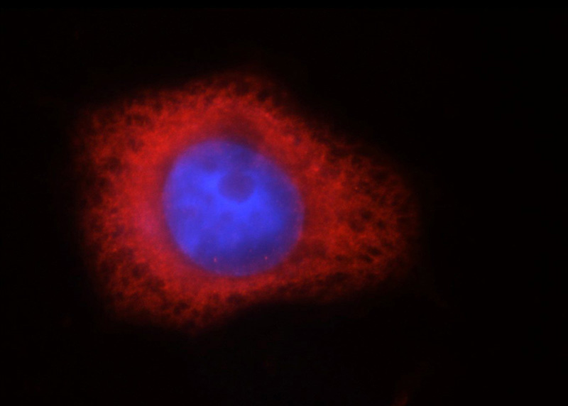
Immunofluorescent analysis of MCF-7 cells, using IGF2BP3 antibody Catalog No:111681 at 1:25 dilution and Rhodamine-labeled goat anti-rabbit IgG (red). Blue pseudocolor = DAPI (fluorescent DNA dye).
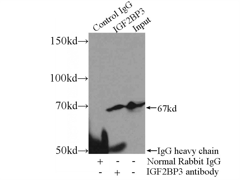
IP Result of anti-IGF2BP3 (IP:Catalog No:111681, 3ug; Detection:Catalog No:111681 1:1000) with HeLa cells lysate 4400ug.
-
Background
IGF2BP3, also named as IMP3, Koc1 and VICKZ3, belongs to the RRM IMP/VICKZ family. It is one of the RNA binding proteins involved in mRNA localization and translational control. IGF2BP3 is expressed during embryogenesis, as well as in some malignant tumors. It can be used as an independent prognostic factor for osteosarcoma. Both isoforms (64kd and 22kd) of IGFBP3 can be recognized by this antibody. And IGFBP3 is nuclear and cytoplasm stains.
-
References
- Li W, Liu D, Chang W. Role of IGF2BP3 in trophoblast cell invasion and migration. Cell death & disease. 5:e1025. 2014.
- Nguyen LH, Robinton DA, Seligson MT. Lin28b is sufficient to drive liver cancer and necessary for its maintenance in murine models. Cancer cell. 26(2):248-61. 2014.
- Wang Y, Yue D, Xiao M. C1QBP negatively regulates the activation of oncoprotein YBX1 in the renal cell carcinoma as revealed by interactomics analysis. Journal of proteome research. 14(2):804-13. 2015.
- Shantha Kumara H, Kirchoff D, Caballero OL. Expression of the cancer testis antigen IGF2BP3 in colorectal cancers; IGF2BP3 holds promise as a specific immunotherapy target. Oncoscience. 2(6):607-14. 2015.
- Chen P, Wang SJ, Wang HB. The distribution of IGF2 and IMP3 in osteosarcoma and its relationship with angiogenesis. Journal of molecular histology. 43(1):63-70. 2012.
- Reschke M, Clohessy JG, Seitzer N. Characterization and analysis of the composition and dynamics of the mammalian riboproteome. Cell reports. 4(6):1276-87. 2013.
Related Products / Services
Please note: All products are "FOR RESEARCH USE ONLY AND ARE NOT INTENDED FOR DIAGNOSTIC OR THERAPEUTIC USE"