-
Product Name
Cytokeratin 7 antibody
- Documents
-
Description
Cytokeratin 7 Rabbit Polyclonal antibody. Positive FC detected in HeLa cells. Positive IHC detected in human ovary tumor tissue, human breast cancer tissue, human liver cancer tissue, human lung cancer tissue, human ovary tissue, human pancreas tissue, human stomach cancer tissue, mouse lung tissue, rat ovary tissue, rat stomach tissue. Positive IF detected in HepG2 cells, HeLa cells. Positive WB detected in HepG2 cells, A431 cells, HeLa cells, mouse liver tissue, rat liver tissue. Observed molecular weight by Western-blot: 54 kDa
-
Tested applications
ELISA, WB, FC, IF, IHC
-
Species reactivity
Human, Mouse, Rat; other species not tested.
-
Alternative names
CK 7 antibody; CK7 antibody; Cytokeratin 7 antibody; K2C7 antibody; keratin 7 antibody; KRT7 antibody; Sarcolectin antibody; SCL antibody; Type II keratin Kb7 antibody
-
Isotype
Rabbit IgG
-
Preparation
This antibody was obtained by immunization of Cytokeratin 7 recombinant protein (Accession Number: NM_005556). Purification method: Antigen affinity purified.
-
Clonality
Polyclonal
-
Formulation
PBS with 0.02% sodium azide and 50% glycerol pH 7.3.
-
Storage instructions
Store at -20℃. DO NOT ALIQUOT
-
Applications
Recommended Dilution:
WB: 1:200-1:2000
IHC: 1:20-1:200
IF: 1:20-1:200
-
Validations
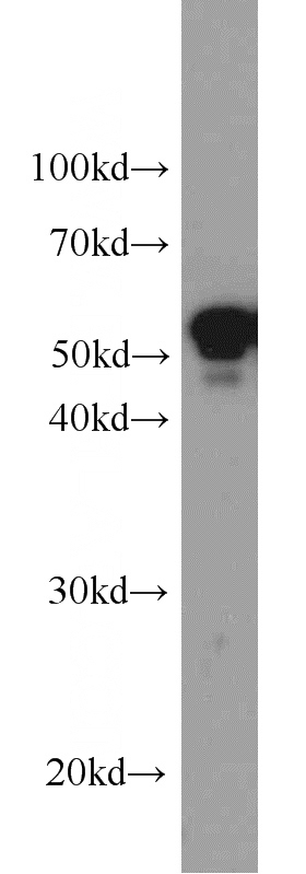
HepG2 cells were subjected to SDS PAGE followed by western blot with Catalog No:109810(KRT7 antibody) at dilution of 1:600
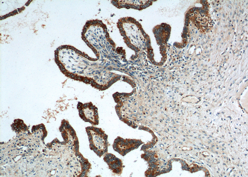
Immunohistochemistry of paraffin-embedded human ovary tumor tissue slide using Catalog No:109810(CK7 Antibody) at dilution of 1:200 (under 10x lens).
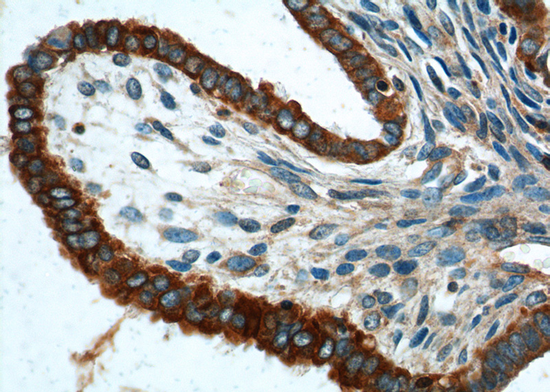
Immunohistochemistry of paraffin-embedded human ovary tumor tissue slide using Catalog No:109810(CK7 Antibody) at dilution of 1:200 (under 40x lens).
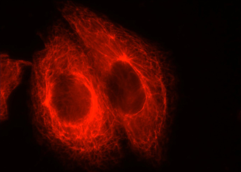
Immunofluorescent analysis of HepG2 cells using Catalog No:109810(KRT7 Antibody) at dilution of 1:50 and Rhodamine-labeled goat anti-rabbit IgG (red).
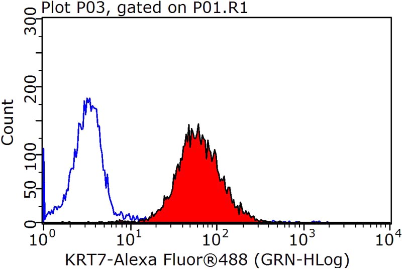
1X10^6 HeLa cells were stained with 0.2ug CK7 antibody (Catalog No:109810, red) and control antibody (blue). Fixed with 90% MeOH blocked with 3% BSA (30 min). Alexa Fluor 488-congugated AffiniPure Goat Anti-Rabbit IgG(H+L) with dilution 1:1500.
-
Background
Keratins are a large family of proteins that form the intermediate filament cytoskeleton of epithelial cells, which are classified into two major sequence types. Type I keratins are a group of acidic intermediate filament proteins, including K9–K23, and the hair keratins Ha1–Ha8. Type II keratins are the basic or neutral courterparts to the acidic type I keratins, including K1–K8, and the hair keratins, Hb1–Hb6. KRT7 is a type II keratin. It is specifically expressed in the simple epithelia lining the cavities of the internal organs and in the gland ducts and blood vessels.
-
References
- Liu S, Wang J, Qin HM. LIF upregulates poFUT1 expression and promotes trophoblast cell migration and invasion at the fetal-maternal interface. Cell death & disease. 5:e1396. 2014.
- Yongping M, Zhang X, Xuewei L. Astragaloside prevents BDL-induced liver fibrosis through inhibition of notch signaling activation. Journal of ethnopharmacology. 169:200-9. 2015.
Related Products / Services
Please note: All products are "FOR RESEARCH USE ONLY AND ARE NOT INTENDED FOR DIAGNOSTIC OR THERAPEUTIC USE"
