
-
Product Name
Anti-IKB alpha (2E10) Mouse antibody
- Documents
-
Description
IKB alpha (2E10) Mouse monoclonal antibody
-
Tested applications
WB
-
Species reactivity
Human, Mouse
-
Alternative names
I kappa B alpha;I kappa B alpha;I(Kappa)B(alpha);I(Kappa)B(alpha);I-kappa-B-alpha; IkappaBalpha;IkB-alpha;IKBA;IKBA;IKBA_HUMAN;IKBalpha;MAD 3;MAD 3;MAD3;Major histocompatibility complex enhancer binding protein MAD3;Major histocompatibility complex enhanc antibody
-
Isotype
Mouse IgG
-
Preparation
Antigen: Purified recombinant human IkB-alpha(N-terminus) protein fragments expressed in E.coli.
-
Clonality
Monoclonal
-
Formulation
Purified mouse monoclonal in buffer containing 0.1M Tris-Glycine (pH 7.4 150 mM NaCl) with 0.02% sodium azide 50% glycerol
-
Storage instructions
Store at 4°C short term. Store at -20°C long term. Avoid freeze / thaw cycle.
-
Applications
WB: 1/1000;Other 1/500
-
Validations
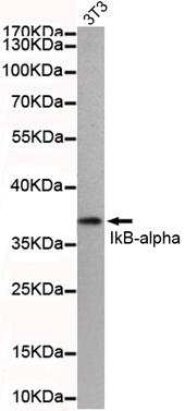
Western blot detection of IkB-alpha(N-terminus) in 3T3 cell lysate using IkB-alpha(N-terminus) mouse mAb (1:500 diluted).Predicted band size: 36KDa.Observed band size: 36KDa.
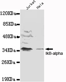
Western blot detection of IkB-alpha(N-terminus) in Jurkat and Hela cell lysates using IkB-alpha(N-terminus) mouse mAb (1:1000 diluted).Predicted band size: 36KDa.Observed band size: 36KDa.
-
Background
Swiss-Prot Acc.P25963.Inhibits the activity of dimeric NF-kappa-B/REL complexes by trapping REL dimers in the cytoplasm through masking of their nuclear localization signals. On cellular stimulation by immune and proinflammatory responses, becomes phosphorylated promoting ubiquitination and degradation, enabling the dimeric RELA to translocate to the nucleus and activate transcription.
Related Products / Services
Please note: All products are "FOR RESEARCH USE ONLY AND ARE NOT INTENDED FOR DIAGNOSTIC OR THERAPEUTIC USE"