
-
Product Name
N-cadherin antibody
- Documents
-
Description
N-cadherin Rabbit Polyclonal antibody. Positive IP detected in mouse brain tissue. Positive WB detected in mouse brain tissue, C6 cells, mouse heart tissue. Positive IHC detected in human brain tissue, human heart tissue. Positive FC detected in SH-SY5Y cells. Observed molecular weight by Western-blot: 130 kDa
-
Tested applications
ELISA, IP, WB, IHC, FC
-
Species reactivity
Human, Mouse, Rat; other species not tested.
-
Alternative names
Cadherin 2 antibody; CD325 antibody; CDH2 antibody; CDHN antibody; CDw325 antibody; N cadherin antibody; NCAD antibody; N-cadherin antibody; Neural cadherin antibody
-
Isotype
Rabbit IgG
-
Preparation
This antibody was obtained by immunization of N-cadherin recombinant protein (Accession Number: NM_001792). Purification method: Antigen affinity purified.
-
Clonality
Polyclonal
-
Formulation
PBS with 0.02% sodium azide and 50% glycerol pH 7.3.
-
Storage instructions
Store at -20℃. DO NOT ALIQUOT
-
Applications
Recommended Dilution:
WB: 1:500-1:5000
IP: 1:500-1:5000
IHC: 1:20-1:200
-
Validations
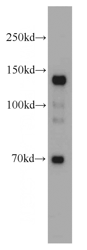
mouse brain tissue were subjected to SDS PAGE followed by western blot with Catalog No:113024(N-cadherin antibody) at dilution of 1:1000
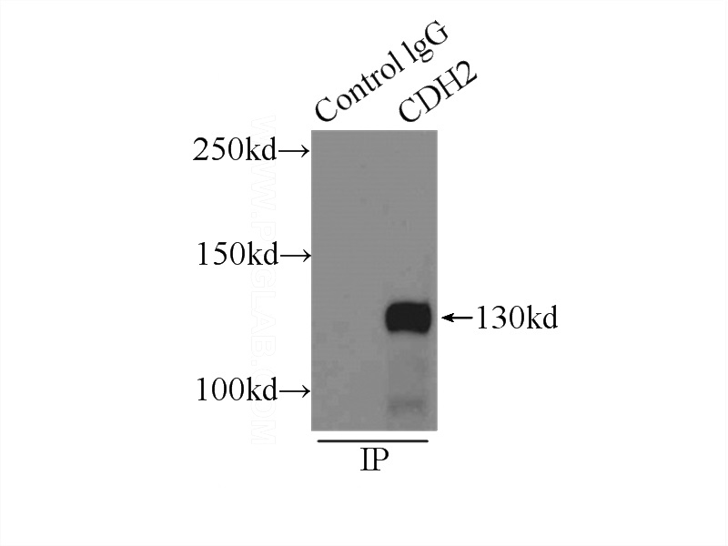
IP Result of anti-N-cadherin (IP:Catalog No:113024, 3ug; Detection:Catalog No:113024 1:1500) with mouse brain tissue lysate 7000ug.
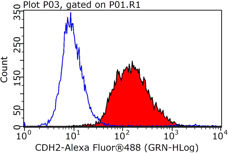
1X10^6 SH-SY5Y cells were stained with 0.2ug N-cadherin antibody (Catalog No:113024, red) and control antibody (blue). Fixed with 90% MeOH blocked with 3% BSA (30 min). Alexa Fluor 488-congugated AffiniPure Goat Anti-Rabbit IgG(H+L) with dilution 1:1000.
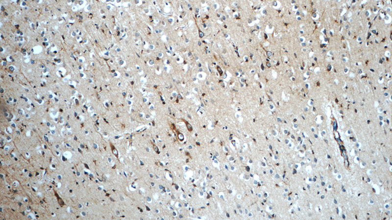
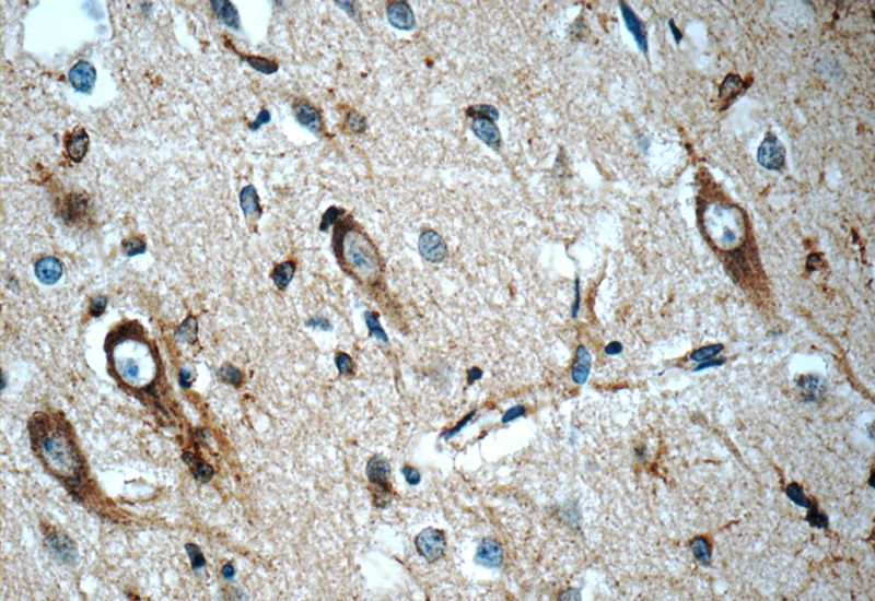
Immunohistochemistry of paraffin-embedded human brain tissue slide using Catalog No:113024(N-cadherin Antibody) at dilution of 1:50 (under 10x lens)
Immunohistochemistry of paraffin-embedded human brain tissue slide using Catalog No:113024(N-cadherin Antibody) at dilution of 1:50 (under 40x lens)
-
Background
Cadherins are a family of transmembrane glycoproteins that mediate calcium-dependent cell-cell adhesion and play an important role in the maintenance of normal tissue architecture. N-cadherin (neural cadherin), also known as CDH2 (cadherin 2), is a classical member of the cadherin superfamily which also include E-, P-, R-, and B-cadherins. Expression of N-cadherin has been reported on various cell types including neurons, endothelial cells and cardiac myocytes (PMID: 11282032; 9508779; 8125202). N-cadherin has functions in early brain morphogenesis, synaptogenesis and synaptic plasticity (PMID: 23321619).
Related Products / Services
Please note: All products are "FOR RESEARCH USE ONLY AND ARE NOT INTENDED FOR DIAGNOSTIC OR THERAPEUTIC USE"