-
Product Name
HDAC2 antibody
- Documents
-
Description
HDAC2 Rabbit Polyclonal antibody. Positive IF detected in HepG2 cells, Hela cells. Positive IHC detected in human prostate cancer tissue, human breast cancer tissue, human testis tissue. Positive FC detected in HEK-293T cells. Positive WB detected in HEK-293 cells, HeLa cells, HepG2 cells, human kidney tissue, Jurkat cells, MCF7 cells, rat liver tissue. Positive IP detected in mouse testis tissue. Observed molecular weight by Western-blot: 55-60 kDa
-
Tested applications
ELISA, WB, IHC, IF, IP, FC
-
Species reactivity
Human,Mouse,Rat; other species not tested.
-
Alternative names
HD2 antibody; HDAC2 antibody; histone deacetylase 2 antibody; RPD3 antibody; YAF1 antibody
-
Isotype
Rabbit IgG
-
Preparation
This antibody was obtained by immunization of HDAC2 recombinant protein (Accession Number: BC031055). Purification method: Antigen affinity purified.
-
Clonality
Polyclonal
-
Formulation
PBS with 0.02% sodium azide and 50% glycerol pH 7.3.
-
Storage instructions
Store at -20℃. DO NOT ALIQUOT
-
Applications
Recommended Dilution:
WB: 1:500-1:5000
IP: 1:500-1:5000
IHC: 1:20-1:200
IF: 1:10-1:100
-
Validations
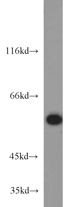
HEK-293 cells were subjected to SDS PAGE followed by western blot with Catalog No:111373(HDAC2 antibody) at dilution of 1:1000
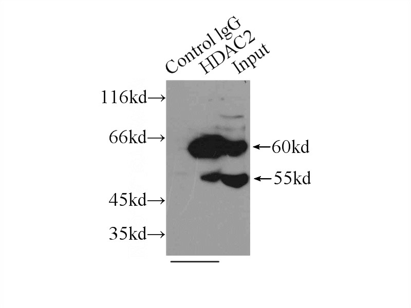
IP Result of anti-HDAC2 (IP:Catalog No:111373, 3ug; Detection:Catalog No:111373 1:1000) with mouse testis tissue lysate 10000ug.
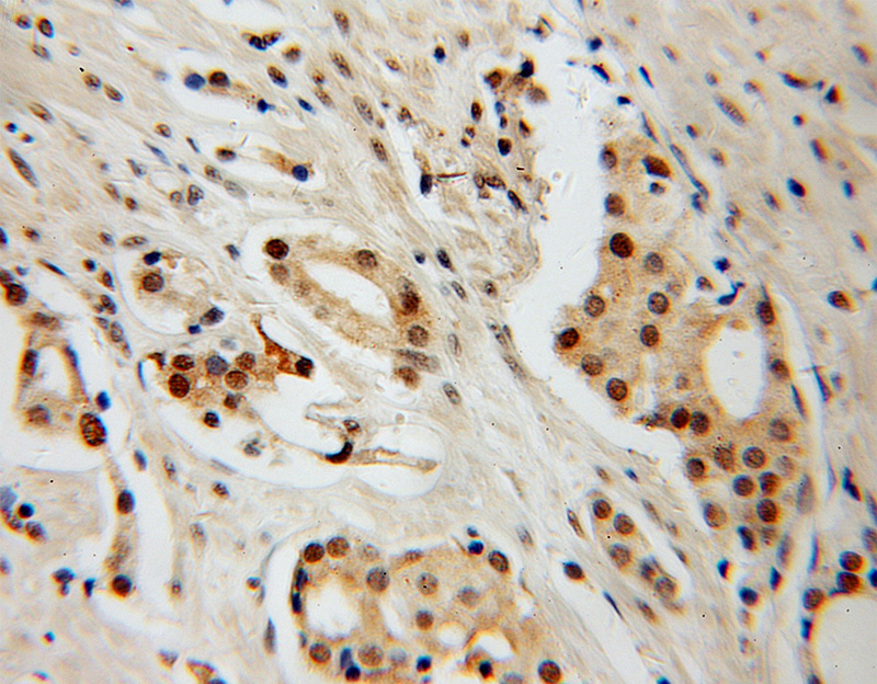
Immunohistochemical of paraffin-embedded human prostate cancer using Catalog No:111373(HDAC2 antibody) at dilution of 1:100 (under 10x lens)
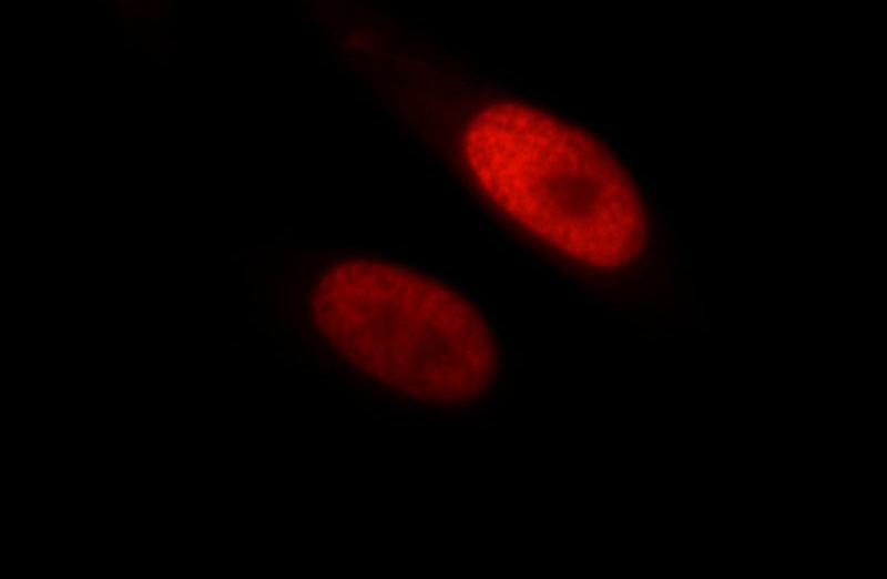
Immunofluorescent analysis of HepG2 cells, using HDAC2 antibody Catalog No:111373 at 1:25 dilution and Rhodamine-labeled goat anti-rabbit IgG (red).
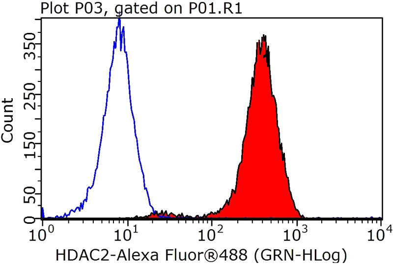
1X10^6 HEK-293T cells were stained with .2ug HDAC2 antibody (Catalog No:111373, red) and control antibody (blue). Fixed with 90% MeOH blocked with 3% BSA (30 min). Alexa Fluor 488-congugated AffiniPure Goat Anti-Rabbit IgG(H+L) with dilution 1:1000.
-
Background
Histone deacetylases(HDAC) are a class of enzymes that remove the acetyl groups from the lysine residues leading to the formation of a condensed and transcriptionally silenced chromatin.Histone deacetylases act via the formation of large multiprotein complexes, and are responsible for the deacetylation of lysine residues at the N-terminal regions of core histones (H2A, H2B, H3 and H4). At least 4 classes of HDAC were identified. As a class I HDAC, HDAC2 was primarily found in the nucleus. HDAC2 forms transcriptional repressor complexes by associating with many different proteins, including YY1, a mammalian zinc-finger transcription factor. Thus, it plays an important role in transcriptional regulation, cell cycle progression and developmental events. This antibody is a rabbit polyclonal antibody raised against residues near the C terminus of human HDAC2.
-
References
- Li Z, Hao Q, Luo J. USP4 inhibits p53 and NF-κB through deubiquitinating and stabilizing HDAC2. Oncogene. 2015.
- Wang X, Liu J, Zhen J. Histone deacetylase 4 selectively contributes to podocyte injury in diabetic nephropathy. Kidney international. 86(4):712-25. 2014.
- Hu X, Lu X, Liu R. Histone cross-talk connects protein phosphatase 1α (PP1α) and histone deacetylase (HDAC) pathways to regulate the functional transition of bromodomain-containing 4 (BRD4) for inducible gene expression. The Journal of biological chemistry. 289(33):23154-67. 2014.
- Wu S, Ge Y, Huang L, Liu H, Xue Y, Zhao Y. BRG1, the ATPase subunit of SWI/SNF chromatin remodeling complex, interacts with HDAC2 to modulate telomerase expression in human cancer cells. Cell cycle (Georgetown, Tex.). 13(18):2869-78. 2014.
- Cai M, Hu Z, Liu J. Expression of hMOF in different ovarian tissues and its effects on ovarian cancer prognosis. Oncology reports. 33(2):685-92. 2015.
Related Products / Services
Please note: All products are "FOR RESEARCH USE ONLY AND ARE NOT INTENDED FOR DIAGNOSTIC OR THERAPEUTIC USE"
