-
Product Name
Cytoglobin antibody
- Documents
-
Description
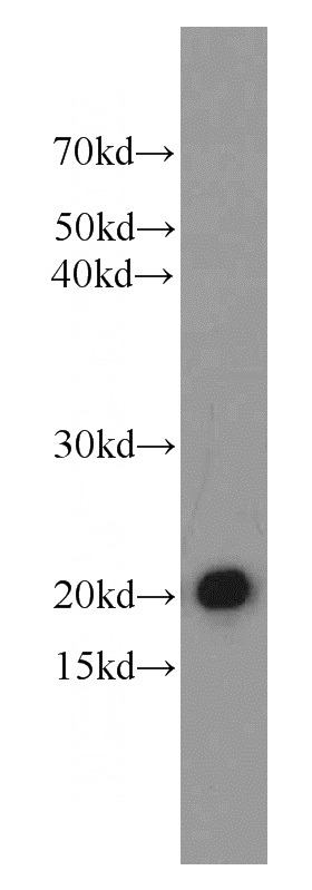
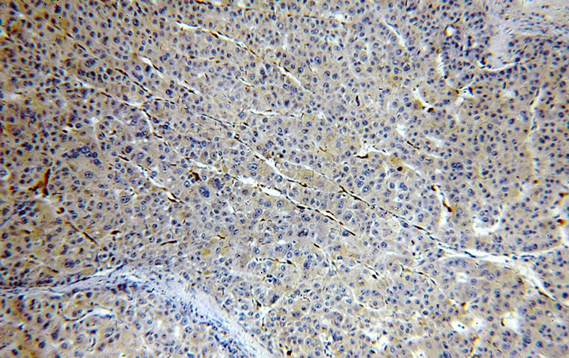
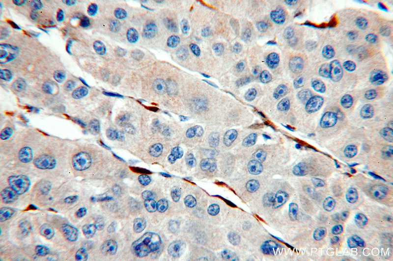
Cytoglobin Rabbit Polyclonal antibody. Positive IHC detected in human liver cancer tissue, human heart tissue, human liver tissue, human prostate cancer tissue, human stomach tissue. Positive WB detected in mouse heart tissue, human heart tissue, human uterus tissue, mouse brain tissue, mouse kidney tissue, mouse lung tissue, mouse small intestine tissue, mouse spleen tissue, mouse stomach tissue, PC-3 cells, rat kidney tissue, Transfected HEK-293 cells. Observed molecular weight by Western-blot: 21-29 kDa
-
Tested applications
ELISA, IHC, WB
-
Species reactivity
Human,Mouse,Rat; other species not tested.
-
Alternative names
CYGB antibody; cytoglobin antibody; HGB antibody; Histoglobin antibody; STAP antibody
-
Isotype
Rabbit IgG
-
Preparation
This antibody was obtained by immunization of Cytoglobin recombinant protein (Accession Number: NM_134268). Purification method: Antigen affinity purified.
-
Clonality
Polyclonal
-
Formulation
PBS with 0.02% sodium azide and 50% glycerol pH 7.3.
-
Storage instructions
Store at -20℃. DO NOT ALIQUOT
-
Applications
Recommended Dilution:
WB: 1:500-1:5000
IHC: 1:20-1:200
-
Validations

mouse heart tissue were subjected to SDS PAGE followed by western blot with Catalog No:109786(CYGB antibody) at dilution of 1:1500

Immunohistochemical of paraffin-embedded human liver cancer using Catalog No:109786(CYGB antibody) at dilution of 1:100 (under 10x lens)

Immunohistochemical of paraffin-embedded human liver cancer using Catalog No:109786(CYGB antibody) at dilution of 1:100 (under 40x lens)
-
Background
Cytoglobin, also named as Histoglobin, HGb, STAP and CYGB, belongs to the globin family. Cytoglobin may have a protective function during conditions of oxidative stress. It may be involved in intracellular oxygen storage or transfer. It serves as a defensive mechanism against oxidative stress both in vitro and in vivo.(PMID:21224051) Cytoglobin is a ubiquitously expressed hexacoordinate hemoglobin that may facilitate diffusion of oxygen through tissues. The protein, with calculated MW of 21 kd, runs as a 29kd one in canine retina, kidney, liver, lung, and heart. It may be caused by posttranslational modification of the protein. Various immuno-staining patterns were reported including nuclear, cytoplasm and extracellular location.
Related Products / Services
Please note: All products are "FOR RESEARCH USE ONLY AND ARE NOT INTENDED FOR DIAGNOSTIC OR THERAPEUTIC USE"
