
-
Product Name
CD31 antibody
- Documents
-
Description
CD31 Rabbit Polyclonal antibody. Positive IHC detected in human renal cell carcinoma tissue, human endometrial cancer tissue, human gliomas tissue, human hepat℃irrhosis tissue, human hysteromyoma tissue, human liver tissue, human lung cancer tissue, human tonsil tissue, human tonsillitis tissue. Positive FC detected in HUVEC cells. Positive IP detected in Jurkat cells. Positive WB detected in Jurkat cells. Observed molecular weight by Western-blot: 80-100 kDa,130 kDa
-
Tested applications
ELISA, IP, IHC, WB, FC
-
Species reactivity
Human; other species not tested.
-
Alternative names
CD31 antibody; EndOCAM antibody; FLJ58394 antibody; GPIIA antibody; PECA1 antibody; PECAM 1 antibody; PECAM1 antibody; VEC marker antibody
-
Isotype
Rabbit IgG
-
Preparation
This antibody was obtained by immunization of CD31 recombinant protein (Accession Number: XM_011524889). Purification method: Antigen affinity purified.
-
Clonality
Polyclonal
-
Formulation
PBS with 0.1% sodium azide and 50% glycerol pH 7.3.
-
Storage instructions
Store at -20℃. DO NOT ALIQUOT
-
Applications
Recommended Dilution:
WB: 1:500-1:5000
IP: 1:200-1:1000
IHC: 1:20-1:200
-
Validations
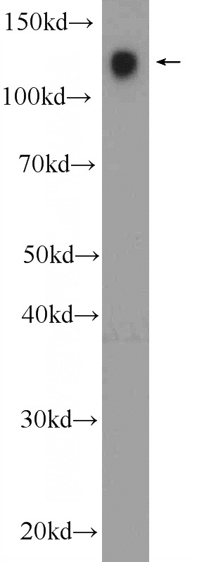
Jurkat cells were subjected to SDS PAGE followed by western blot with Catalog No:109025(CD31 Antibody) at dilution of 1:1000
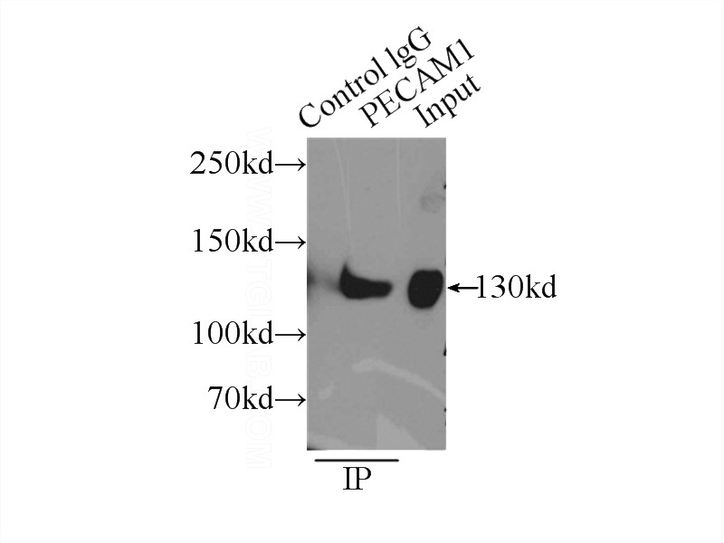
IP Result of anti-PECAM1,CD31 (IP:Catalog No:109025, 5ug; Detection:Catalog No:109025 1:300) with Jurkat cells lysate 4000ug.
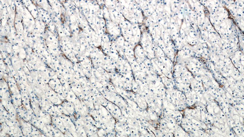
Immunohistochemistry of paraffin-embedded human renal cell carcinoma tissue slide using Catalog No:109025(CD31 Antibody) at dilution of 1:400 (under 10x lens)
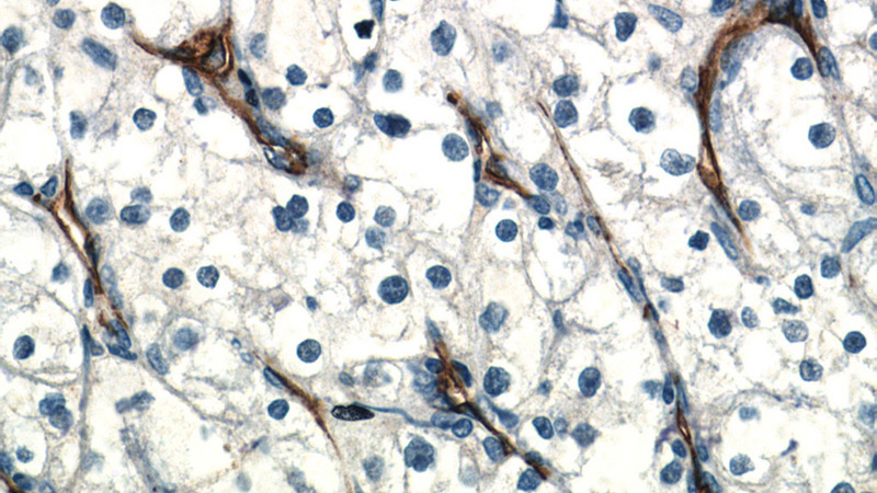
Immunohistochemistry of paraffin-embedded human renal cell carcinoma tissue slide using Catalog No:109025(CD31 Antibody) at dilution of 1:400 (under 40x lens)
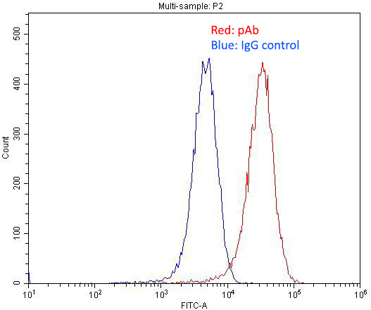
1X10^6 HUVEC cells were stained with 0.2ug CD31 antibody (Catalog No:109025, red) and control antibody (blue). Fixed with 4% PFA blocked with 3% BSA (30 min). Alexa Fluor 488-congugated AffiniPure Goat Anti-Rabbit IgG(H+L) with dilution 1:1500.
-
Background
Platelet endothelial cell adhesion molecule-1 (PECAM-1, CD31) is a member of the immunoglobulin gene superfamily of cell adhesion molecules. It is highly expressed on the surface of the endothelium, making up a large portion of its intracellular junctions. PECAM-1 is also present on the surface of hematopoietic cells and immune cells including platelets, monocytes, neutrophils, natural killer cells, megakaryocytes and some types of T-cell (PMID: 9011572). As well as its role in cell-cell adhesion, PECAM-1 functions as a signaling receptor, and is involved in important physiological events such as nitric oxide production, regulation of T-cell immunity and tolerance, leukocyte transendothelial migration and inflammation and angiogenesis (PMID: 21183735; 20978210; 17872453; 20634489).
-
References
- Lu SJ, Ivanova Y, Feng Q, Luo C, Lanza R. Hemangioblasts from human embryonic stem cells generate multilayered blood vessels with functional smooth muscle cells. Regenerative medicine. 4(1):37-47. 2009.
- Okamoto H, Umeda S, Nozawa T. Comparative proteomic analyses of macular and peripheral retina of cynomolgus monkeys (Macaca fascicularis). Experimental animals / Japanese Association for Laboratory Animal Science. 59(2):171-82. 2010.
- Zhou B, Ma R, Si W. MicroRNA-503 targets FGF2 and VEGFA and inhibits tumor angiogenesis and growth. Cancer letters. 333(2):159-69. 2013.
- Rossdeutsch A, Smart N, Dubé KN, Turner M, Riley PR. Essential role for thymosin β4 in regulating vascular smooth muscle cell development and vessel wall stability. Circulation research. 111(4):e89-102. 2012.
- Wang HL, Wang SS, Song WH. Expression of prostate-specific membrane antigen in lung cancer cells and tumor neovasculature endothelial cells and its clinical significance. PloS one. 10(5):e0125924. 2015.
- Chen K, Yu G, Gumireddy K. ZBRK1, a novel tumor suppressor, activates VHL gene transcription through formation of a complex with VHL and p300 in renal cancer. Oncotarget. 6(9):6959-76. 2015.
- Zhou ZJ, Dai Z, Zhou SL. HNRNPAB induces epithelial-mesenchymal transition and promotes metastasis of hepatocellular carcinoma by transcriptionally activating SNAIL. Cancer research. 74(10):2750-62. 2014.
- Vishnubalaji R, Atteya M, Al-Nbaheen M, Oreffo RO, Aldahmash A, Alajez NM. Angiogenic Potential of Human Neonatal Foreskin Stromal Cells in the Chick Embryo Chorioallantoic Membrane Model. Stem cells international. 2015:257019. 2015.
Related Products / Services
Please note: All products are "FOR RESEARCH USE ONLY AND ARE NOT INTENDED FOR DIAGNOSTIC OR THERAPEUTIC USE"