
-
Product Name
Anti-PI3 Kinase p85 alpha (9D2) Mouse antibody
- Documents
-
Description
PI3 Kinase p85 alpha (9D2) Mouse monoclonal antibody
-
Tested applications
IHC-P, ICC/IF, FC
-
Species reactivity
Human, Mouse
-
Isotype
Mouse IgG1
-
Preparation
Antigen: Purified recombinant fragment of human PIK3R1 (AA: 159-388) expressed in E. Coli.
-
Clonality
Monoclonal
-
Formulation
Purified antibody in PBS with 0.05% sodium azide
-
Storage instructions
Store at 4°C short term. Store at -20°C long term. Avoid freeze / thaw cycle.
-
Applications
IHC: 1/200 - 1/1000
ICC: 1/100
FC: 1/200 - 1/400
ELISA: 1/10000
-
Validations
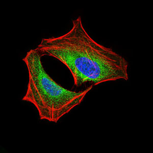
Immunofluorescence analysis of HeLa cells using PIK3R1 mouse mAb (green). Blue: DRAQ5 fluorescent DNA dye. Red: Actin filaments have been labeled with Alexa Fluor-555 phalloidin.
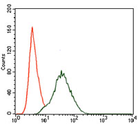
Flow cytometric analysis of NIH3T3 cells using PIK3R1 mouse mAb (green) and negative control (red).
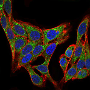
Immunofluorescence analysis of HepG2 cells using PIK3R1 mouse mAb (green). Blue: DRAQ5 fluorescent DNA dye. Red: Actin filaments have been labeled with Alexa Fluor-555 phalloidin.
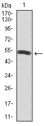
Western blot analysis using PIK3R1 mAb against human PIK3R1 recombinant protein. (Expected MW is 53.4 kDa)
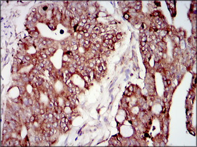
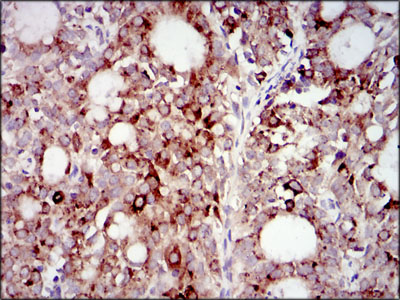
Immunohistochemical analysis of paraffin-embedded rectum cancer tissues using PIK3R1 mouse mAb with DAB staining.
Immunohistochemical analysis of paraffin-embedded cervical cancer tissues using PIK3R1 mouse mAb with DAB staining.
-
Background
Swiss-Prot Acc.P27986.Phosphatidylinositol 3-kinase phosphorylates the inositol ring of phosphatidylinositol at the 3-prime position. The enzyme comprises a 110 kD catalytic subunit and a regulatory subunit of either 85, 55, or 50 kD. This gene encodes the 85 kD regulatory subunit. Phosphatidylinositol 3-kinase plays an important role in the metabolic actions of insulin, and a mutation in this gene has been associated with insulin resistance. Alternative splicing of this gene results in four transcript variants encoding different isoforms.
Related Products / Services
Please note: All products are "FOR RESEARCH USE ONLY AND ARE NOT INTENDED FOR DIAGNOSTIC OR THERAPEUTIC USE"