
-
Product Name
Anti-Anti-CD44 Mouse antibody
- Documents
-
Description
Anti-CD44 Mouse monoclonal antibody
-
Tested applications
IHC-P, WB, ICC/IF, FC
-
Species reactivity
Human
-
Isotype
Mouse IgG
-
Preparation
Antigen: This CD44 monoclonal antibody is generated from mouse immunized with CD44 recombinant protein.
-
Clonality
Monoclonal
-
Formulation
Purified monoclonal antibody supplied in PBS with 0.09% (W/V) sodium azide. This antibody is purified through a protein G column, eluted with high and low pH buffers and neutralized immediately, followed by dialysis against PBS.
-
Applications
WB::1:2000ICC::1:10~50
-
Validations
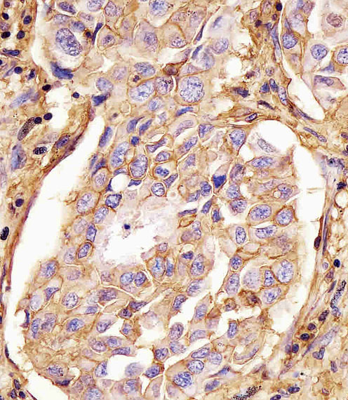
169110 staining CD44 in human lung adenocarcinoma tissue sections by Immunohistochemistry (IHC-P - paraformaldehyde-fixed, paraffin-embedded sections). Tissue was fixed with formaldehyde and blocked with 3% BSA for 0. 5 hour at room temperature; antigen retrieval was by heat mediation with a citrate buffer (pH6). Samples were incubated with primary antibody (1/25) for 1 hours at 37°C. A undiluted biotinylated goat polyvalent antibody was used as the secondary antibody.
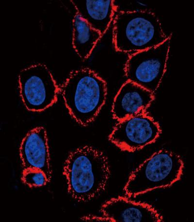
CD44 antibody (Cat. #169110) confocal immunofluorescent analysis with hela cell. 0.01 mg/ml primary antibody was followed by PE-conjugated goat anti-mouse lgG (whole molecule). PE emits red fluorescence. DAPI was used to stain the cell nuclear (blue).
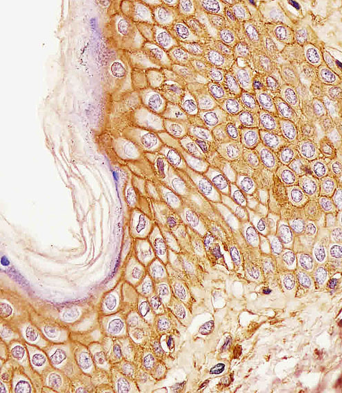
169110 staining CD44 in human skin tissue sections by Immunohistochemistry (IHC-P - paraformaldehyde-fixed, paraffin-embedded sections). Tissue was fixed with formaldehyde and blocked with 3% BSA for 0. 5 hour at room temperature; antigen retrieval was by heat mediation with a citrate buffer (pH6). Samples were incubated with primary antibody (1/25) for 1 hours at 37°C. A undiluted biotinylated goat polyvalent antibody was used as the secondary antibody.
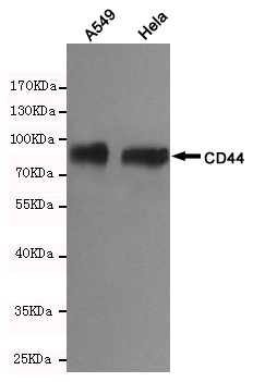
Western blot detection of CD44 in A549 and Hela cells using CD44 Mouse mAb(dilution 1:1000).Predicted band size:81kDa.Observed band size:81kDa.
-
Background
Swiss-Prot Acc.P16070.
Related Products / Services
Please note: All products are "FOR RESEARCH USE ONLY AND ARE NOT INTENDED FOR DIAGNOSTIC OR THERAPEUTIC USE"