
-
Product Name
TDP-43 (C-terminal) antibody
- Documents
-
Description
TDP-43 (C-terminal) Rabbit Polyclonal antibody. Positive IP detected in HeLa cells, mouse brain tissue. Positive WB detected in HeLa cells, A2780 cells, A549 cells, C6 cells, human heart tissue, human placenta tissue, K-562 cells, mouse heart tissue, mouse pancreas tissue. Positive IF detected in A549 cells, HeLa cells. Positive IHC detected in human lung cancer tissue, human gliomas tissue, mouse brain tissue, mouse pancreas tissue. Observed molecular weight by Western-blot: 43-45 kDa,35 kDa
-
Tested applications
ELISA, IHC, IF, IP, WB
-
Species reactivity
Human,Mouse,Rat; other species not tested.
-
Alternative names
ALS10 antibody; TAR DNA binding protein antibody; TAR DNA binding protein 43 antibody; TARDBP antibody; TDP 43 antibody; TDP43 antibody
-
Isotype
Rabbit IgG
-
Preparation
This antibody was obtained by immunization of TDP-43 (C-terminal) recombinant protein (Accession Number: BC001487). Purification method: Antigen affinity purified.
-
Clonality
Polyclonal
-
Formulation
PBS with 0.1% sodium azide and 50% glycerol pH 7.3.
-
Storage instructions
Store at -20℃. DO NOT ALIQUOT
-
Applications
Recommended Dilution:
WB: 1:500-1:5000
IP: 1:500-1:5000
IHC: 1:20-1:200
IF: 1:20-1:200
-
Validations
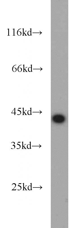
HeLa cells were subjected to SDS PAGE followed by western blot with Catalog No:115926(TARDBP antibody) at dilution of 1:1000
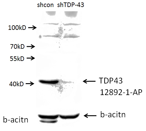
A549 cells (shcontrol and shRNA of TDP43) were subjected to SDS PAGE followed by western blot with Catalog No:115926 (TDP43 antibody) at dilution of 1:1000. (Data provided by Angran Biotech (www.miRNAlab.com)).
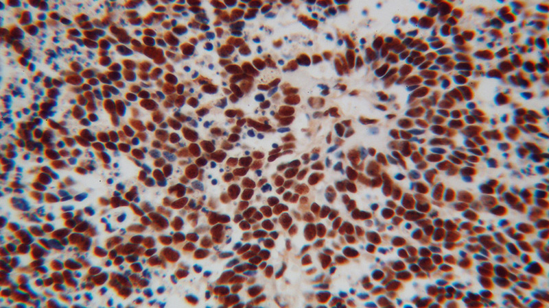
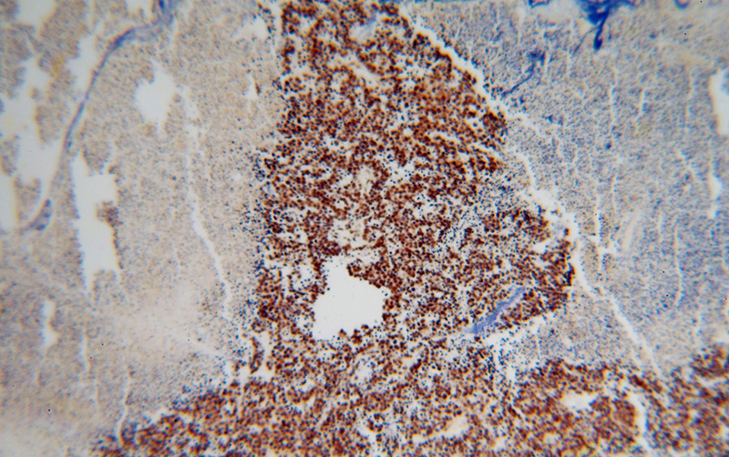
Immunohistochemical of paraffin-embedded human lung cancer using Catalog No:115926(TARDBP antibody) at dilution of 1:100 (under 40x lens)
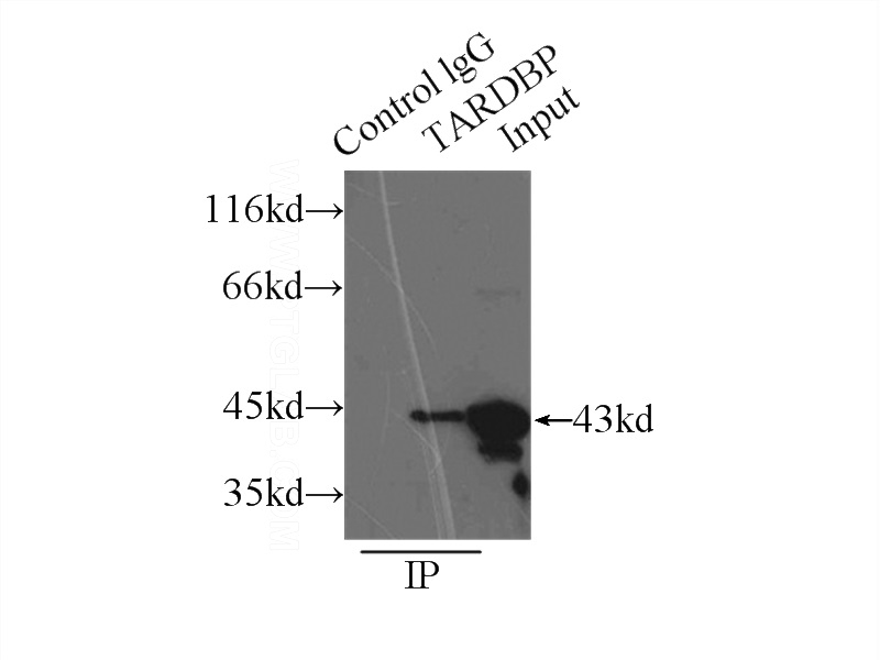
IP Result of anti-TARDBP (IP:Catalog No:115926, 3ug; Detection:Catalog No:115926 1:1000) with HeLa cells lysate 3000ug.
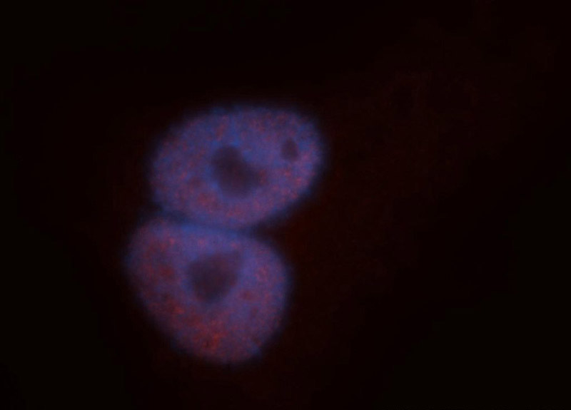
Immunofluorescent analysis of A549 cells, using TARDBP antibody Catalog No:115926 at 1:50 dilution and Rhodamine-labeled goat anti-rabbit IgG (red). Blue pseudocolor = DAPI (fluorescent DNA dye).
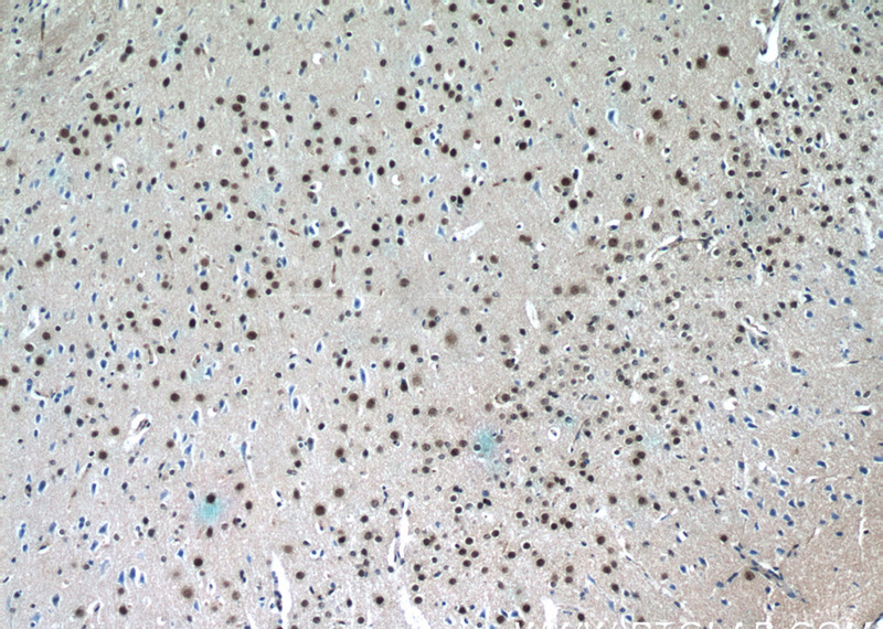
Immunohistochemistry of paraffin-embedded mouse brain tissue slide using Catalog No:115926(TDP-43 Antibody) at dilution of 1:50 (under 10x lens)
Immunohistochemical of paraffin-embedded human lung cancer using Catalog No:115926(TARDBP antibody) at dilution of 1:100 (under 10x lens)
-
Background
Transactivation response (TAR) DNA-binding protein of 43 kDa (also known as TARDBP or TDP-43) was first isolated as a transcriptional inactivator binding to the TAR DNA element of the HIV-1 virus. Neumann et al. (2006) found that a hyperphosphorylated, ubiquitinated, and cleaved form of TARDBP, known as pathologic TDP-43, is the major component of the tau-negative and ubiquitin-positive inclusions that characterize amyotrophic lateral sclerosis (ALS) and the most common pathological subtype of frontotemporal lobar degeneration (FTLD-U). 12892-1-AP is a rabbit polyclonal antibody raised against the C-terminal amino acids of human TDP-43. This antibody recognizes the cleavage product of 20-30 kDa in addition to the native and phosphorylated forms of TDP-43. Immunohistochemical analyses of TDP-43 using this antibody detect both normal diffuse nuclear staining and insoluble inclusions in pathologic tissues.
-
References
- Suzuki H, Shibagaki Y, Hattori S, Matsuoka M. Nuclear TDP-43 causes neuronal toxicity by escaping from the inhibitory regulation by hnRNPs. Human molecular genetics. 24(6):1513-27. 2015.
- Saldi TK, Ash PE, Wilson G. TDP-1, the Caenorhabditis elegans ortholog of TDP-43, limits the accumulation of double-stranded RNA. The EMBO journal. 33(24):2947-66. 2014.
- Han H, Wei W, Duan W. Autophagy-linked FYVE protein (Alfy) promotes autophagic removal of misfolded proteins involved in amyotrophic lateral sclerosis (ALS). In vitro cellular & developmental biology. Animal. 51(3):249-63. 2015.
- Suzuki H, Matsuoka M. Overexpression of nuclear FUS induces neuronal cell death. Neuroscience. 287:113-24. 2015.
- Raitano S, Ordovàs L, De Muynck L. Restoration of progranulin expression rescues cortical neuron generation in an induced pluripotent stem cell model of frontotemporal dementia. Stem cell reports. 4(1):16-24. 2015.
- Paré B, Touzel-Deschênes L, Lamontagne R. Early detection of structural abnormalities and cytoplasmic accumulation of TDP-43 in tissue-engineered skins derived from ALS patients. Acta neuropathologica communications. 3:5. 2015.
- Goossens J, Vanmechelen E, Trojanowski JQ. TDP-43 as a possible biomarker for frontotemporal lobar degeneration: a systematic review of existing antibodies. Acta neuropathologica communications. 3:15. 2015.
- Huang CC, Bose JK, Majumder P. Metabolism and mis-metabolism of the neuropathological signature protein TDP-43. Journal of cell science. 127(Pt 14):3024-38. 2014.
Related Products / Services
Please note: All products are "FOR RESEARCH USE ONLY AND ARE NOT INTENDED FOR DIAGNOSTIC OR THERAPEUTIC USE"