-
Product Name
Stathmin 1 antibody
- Documents
-
Description
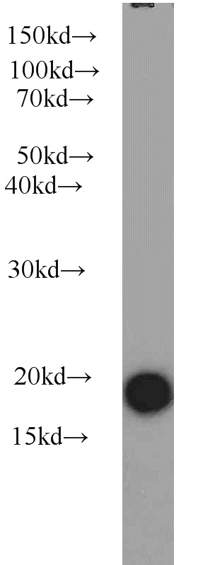
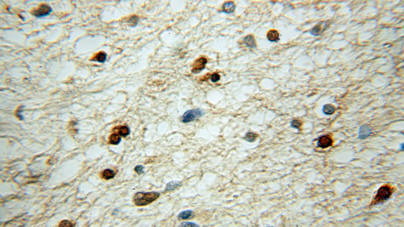
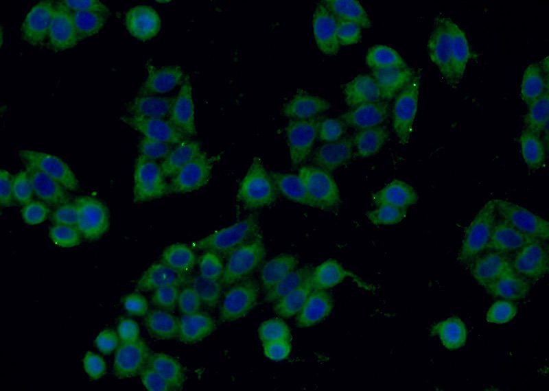
Stathmin 1 Rabbit Polyclonal antibody. Positive WB detected in Jurkat cells, HeLa cells, human brain tissue, K-562 cells, MDA-MB-453s cells, mouse brain tissue, rat brain tissue. Positive IHC detected in human gliomas tissue, human lymphoma tissue. Positive IF detected in HeLa cells. Observed molecular weight by Western-blot: 18 kDa
-
Tested applications
ELISA, WB, IHC, IF
-
Species reactivity
Human,Mouse,Rat; other species not tested.
-
Alternative names
C1orf215 antibody; FLJ32206 antibody; Lag antibody; LAP18 antibody; Metablastin antibody; Oncoprotein 18 antibody; OP18 antibody; Phosphoprotein p19 antibody; PP17 antibody; PP19 antibody; PR22 antibody; Prosolin antibody; Protein Pr22 antibody; SMN antibody; Stathmin antibody; stathmin 1/oncoprotein 18 antibody; Stathmin1 antibody; STMN1 antibody
-
Isotype
Rabbit IgG
-
Preparation
This antibody was obtained by immunization of Stathmin 1 recombinant protein (Accession Number: NM_203399). Purification method: Antigen affinity purified.
-
Clonality
Polyclonal
-
Formulation
PBS with 0.1% sodium azide and 50% glycerol pH 7.3.
-
Storage instructions
Store at -20℃. DO NOT ALIQUOT
-
Applications
Recommended Dilution:
WB: 1:200-1:2000
IHC: 1:20-1:200
IF: 1:20-1:200
-
Validations

Jurkat cells were subjected to SDS PAGE followed by western blot with Catalog No:115698(STMN1 antibody) at dilution of 1:800

Immunohistochemical of paraffin-embedded human gliomas using Catalog No:115698(STMN1 antibody) at dilution of 1:50 (under 10x lens)

Immunofluorescent analysis of HeLa cells using Catalog No:115698(STMN1 Antibody) at dilution of 1:50 and Alexa Fluor 488-congugated AffiniPure Goat Anti-Rabbit IgG(H+L)
-
Background
Stathmin 1 (STMN1) normally regulates microtubule dynamics either by sequestering free tubulin heterodimers or by promoting microtubule catastrophe. STMN1 is highly expressed in fetal and adult brain, spinal cord, and cerebellum. Many different phosphorylated forms are observed depending on specific combinations among the sites which can be phosphorylated. Phosphorylation of stathmin is involved in response to NGF, neuron polarization and microtubule polymerization inhibition activity. Increased expression of STMN1 has been observed in a variety of human malignancies, such as colorectal primary tumors and metastatic tissues, but its association with melanoma is so far not well known.
-
References
- Zhang Y, Xiong J, Wang J. Regulation of melanocyte apoptosis by Stathmin 1 expression. BMB reports. 41(11):765-70. 2008.
- Ma D, Cui L, Gao J. Proteomic analysis of mesenchymal stem cells from normal and deep carious dental pulp. PloS one. 9(5):e97026. 2014.
- Zhang L, Feng D, Tao H, DE X, Chang Q, Hu Q. Increased stathmin expression strengthens fear conditioning in epileptic rats. Biomedical reports. 3(1):28-32. 2015.
- Fuller HR, Slade R, Jovanov-Milošević N. Stathmin is enriched in the developing corticospinal tract. Molecular and cellular neurosciences. 69:12-21. 2015.
- Kuang XY, Chen L, Zhang ZJ. Stathmin and phospho-stathmin protein signature is associated with survival outcomes of breast cancer patients. Oncotarget. 6(26):22227-38. 2015.
Related Products / Services
Please note: All products are "FOR RESEARCH USE ONLY AND ARE NOT INTENDED FOR DIAGNOSTIC OR THERAPEUTIC USE"
