-
Product Name
SMCR7L/MID51 antibody
- Documents
-
Description
SMCR7L/MID51 Rabbit Polyclonal antibody. Positive WB detected in HT-1080 cells, mouse heart tissue, mouse testis tissue, NIH/3T3 cells, rat liver tissue, rat testis tissue, RAW264.7 cells. Positive IP detected in RAW 264.7 cells. Positive IF detected in Hela cells. Observed molecular weight by Western-blot: 48-51 kDa
-
Tested applications
ELISA, WB, IF, IP
-
Species reactivity
Human, Mouse, Rat; other species not tested.
-
Alternative names
dJ1104E15.3 antibody; FLJ20232 antibody; HSU79252 antibody; MID51 antibody; MIEF1 antibody; SMCR7L antibody
- Immunogen
-
Isotype
Rabbit IgG
-
Preparation
This antibody was obtained by immunization of SMCR7L/MID51 recombinant protein (Accession Number: BC002587). Purification method: Antigen affinity purified.
-
Clonality
Polyclonal
-
Formulation
PBS with 0.02% sodium azide and 50% glycerol pH 7.3.
-
Storage instructions
Store at -20℃. DO NOT ALIQUOT
-
Applications
Recommended Dilution:
WB: 1:500-1:5000
IP: 1:500-1:5000
IF: 1:10-1:100
-
Validations

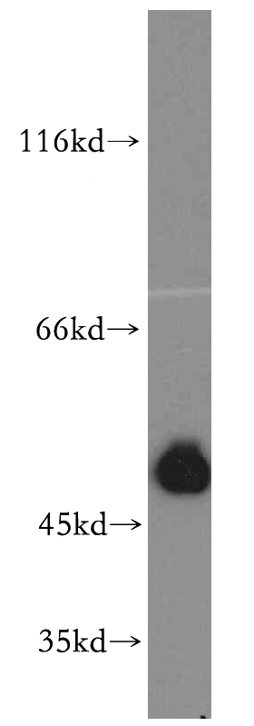
HT-1080 cells were subjected to SDS PAGE followed by western blot with Catalog No:115387(SMCR7L antibody) at dilution of 1:400

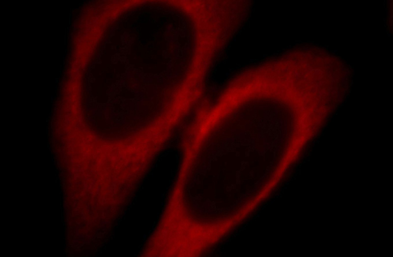
Immunofluorescent analysis of Hela cells, using SMCR7L antibody Catalog No:115387 at 1:25 dilution and Rhodamine-labeled goat anti-rabbit IgG (red).

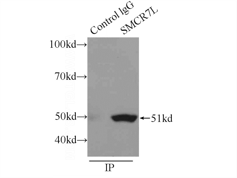
IP Result of anti-SMCR7L (IP:Catalog No:115387, 3ug; Detection:Catalog No:115387 1:1000) with RAW 264.7 cells lysate 2400ug.
-
Background
Human SMCR7L gene encodes, MID51, the mitochondrial dynamic protein of 51 kDa (also called mitochondrial elongation factor 1, MIEF1). MID51 is a single-pass membrane protein anchored to the mitochondrial outer membrane and regulates mitochondrial morphology. Mitochondrial morphology is controlled by two opposing processes: fusion and fission. Elevated MID51 levels induce extensive mitochondrial fusion, whereas depletion of MID51 causes mitochondrial fragmentation. MID51 interacts with and recruits Drp1 to mitochondria, suggesting a critical role of MID51 in regulation of mitochondrial fusion-fission machinery in vertebrates.
-
References
- Qi X, Qvit N, Su YC, Mochly-Rosen D. A novel Drp1 inhibitor diminishes aberrant mitochondrial fission and neurotoxicity. Journal of cell science. 126(Pt 3):789-802. 2013.
- Losón OC, Song Z, Chen H, Chan DC. Fis1, Mff, MiD49, and MiD51 mediate Drp1 recruitment in mitochondrial fission. Molecular biology of the cell. 24(5):659-67. 2013.
- Richter V, Palmer CS, Osellame LD. Structural and functional analysis of MiD51, a dynamin receptor required for mitochondrial fission. The Journal of cell biology. 204(4):477-86. 2014.
- Ruan Y, Li H, Zhang K, Jian F, Tang J, Song Z. Loss of Yme1L perturbates mitochondrial dynamics. Cell death & disease. 4:e896. 2013.
- Zhang Z, Liu L, Wu S, Xing D. Drp1, Mff, Fis1, and MiD51 are coordinated to mediate mitochondrial fission during UV irradiation-induced apoptosis. FASEB journal : official publication of the Federation of American Societies for Experimental Biology. 30(1):466-76. 2016.
- Li H, Ruan Y, Zhang K. Mic60/Mitofilin determines MICOS assembly essential for mitochondrial dynamics and mtDNA nucleoid organization. Cell death and differentiation. 23(3):380-92. 2016.
- Xu S, Cherok E, Das S. Mitochondrial E3 ubiquitin ligase MARCH5 controls mitochondrial fission and cell sensitivity to stress-induced apoptosis through regulation of MiD49 protein. Molecular biology of the cell. 27(2):349-59. 2016.
- Vannuvel K, Van Steenbrugge M, Demazy C. Effects of a Sublethal and Transient Stress of the Endoplasmic Reticulum on the Mitochondrial Population. Journal of cellular physiology. 2015.
Related Products / Services
Please note: All products are "FOR RESEARCH USE ONLY AND ARE NOT INTENDED FOR DIAGNOSTIC OR THERAPEUTIC USE"
