-
Product Name
PAK2 antibody
- Documents
-
Description
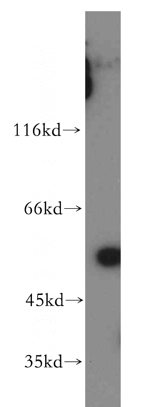
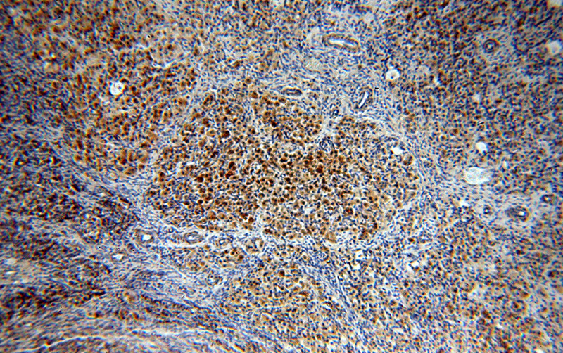
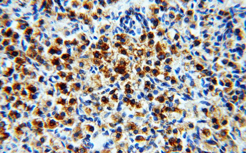
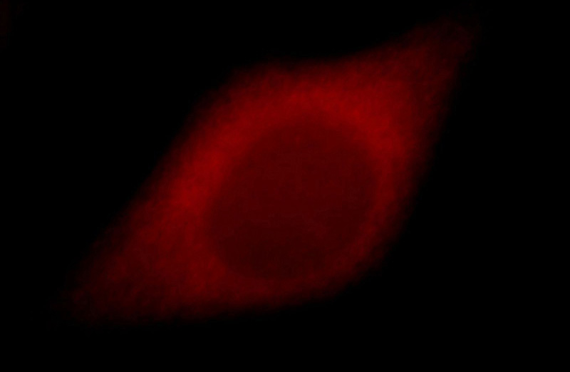
PAK2 Rabbit Polyclonal antibody. Positive IF detected in Hela cells. Positive IHC detected in human ovary tissue, human spleen tissue. Positive WB detected in human skeletal muscle tissue. Observed molecular weight by Western-blot: 54 kDa
-
Tested applications
ELISA, IHC, IF, WB
-
Species reactivity
Human; other species not tested.
-
Alternative names
C t PAK2 antibody; Gamma PAK antibody; p21 activated kinase 2 antibody; p27 PAK 2p34 antibody; p34 antibody; p58 antibody; PAK 2 antibody; PAK2 antibody; PAK65 antibody; PAKgamma antibody; S6/H4 kinase antibody
-
Isotype
Rabbit IgG
-
Preparation
This antibody was obtained by immunization of Peptide (Accession Number: NM_002577). Purification method: Antigen affinity purified.
-
Clonality
Polyclonal
-
Formulation
PBS with 0.02% sodium azide and 50% glycerol pH 7.3.
-
Storage instructions
Store at -20℃. DO NOT ALIQUOT
-
Applications
Recommended Dilution:
WB: 1:200-1:1000
IHC: 1:20-1:200
IF: 1:10-1:100
-
Validations

human skeletal muscle tissue were subjected to SDS PAGE followed by western blot with Catalog No:113499(PAK2 antibody) at dilution of 1:300

Immunohistochemical of paraffin-embedded human ovary using Catalog No:113499(PAK2 antibody) at dilution of 1:100 (under 10x lens)

Immunohistochemical of paraffin-embedded human ovary using Catalog No:113499(PAK2 antibody) at dilution of 1:100 (under 40x lens)

Immunofluorescent analysis of Hela cells, using PAK2 antibody Catalog No:113499 at 1:25 dilution and Rhodamine-labeled goat anti-rabbit IgG (red).
-
Background
PAK2, also named as PAK65, PAKgamma, p58, PAK-2p27, PAK-2p24 and C-t-PAK2, belongs to the protein kinase superfamily, STE Ser/Thr protein kinase family and STE20 subfamily. Full length PAK 2 stimulates cell survival and cell growth. The process is, at least in part, mediated by phosphorylation and inhibition of pro-apoptotic BAD. PAK2 has several isoforms with the MW of 54-62 kDa and 41 kDa. The 62 kDa PAK2 is cleaved into a 34 kDa C terminal fragment and a 28 kDa N terminal fragment with a time course that parallels apoptotic death in Jurkat cells. (PMID:10200518). Caspase-activated PAK-2p34 is involved in cell death response, probably involving the JNK signaling pathway. Cleaved PAK-2p34 seems to have a higher activity than the CDC42-activated form. The antibody has no cross reaction to PAK1 and PAK3.
Related Products / Services
Please note: All products are "FOR RESEARCH USE ONLY AND ARE NOT INTENDED FOR DIAGNOSTIC OR THERAPEUTIC USE"
