
-
Product Name
P16-INK4A antibody
- Documents
-
Description
P16-INK4A Rabbit Polyclonal antibody. Positive IF detected in SH-SY5Y cells. Positive IHC detected in human lung cancer tissue, human cervical cancer tissue, human lymphoma tissue. Positive FC detected in HeLa cells. Positive IP detected in HEK-293 cells. Positive WB detected in HEK-293 cells, HeLa cells, PC-3 cells. Observed molecular weight by Western-blot: 16-18 kDa
-
Tested applications
ELISA, WB, IF, IP, FC, IHC
-
Species reactivity
Human; other species not tested.
-
Alternative names
ARF antibody; ARF antibody; CDKN2A antibody; CDK4 inhibitor p16 INK4 antibody; CDK4 inhibitor p16INK4 antibody; CDK4I antibody; CDKN2 antibody; CDKN2A antibody; CMM2 antibody; INK4 antibody; INK4a antibody; MLM antibody; MTS 1 antibody; MTS1 antibody; Multiple tumor suppressor 1 antibody; p14 antibody; p14ARF antibody; P16 antibody; p16 INK4 antibody; p16 INK4a antibody; p16INK4 antibody; p16INK4a antibody; P19 antibody; TP16 antibody
-
Isotype
Rabbit IgG
-
Preparation
This antibody was obtained by immunization of P16-INK4A recombinant protein (Accession Number: BC021998). Purification method: Antigen affinity purified.
-
Clonality
Polyclonal
-
Formulation
PBS with 0.1% sodium azide and 50% glycerol pH 7.3.
-
Storage instructions
Store at -20℃. DO NOT ALIQUOT
-
Applications
Recommended Dilution:
WB: 1:500-1:5000
IP: 1:500-1:5000
IHC: 1:50-1:500
IF: 1:20-1:200
-
Validations
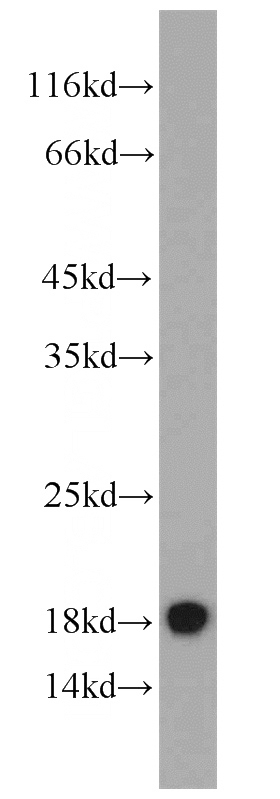
HEK-293 cells were subjected to SDS PAGE followed by western blot with Catalog No:113538(P16 antibody) at dilution of 1:1000
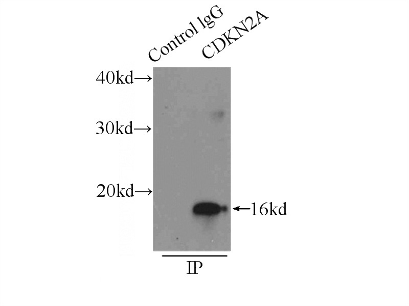
IP Result of anti-P16 (IP:Catalog No:113538, 4ug; Detection:Catalog No:113538 1:1000) with HEK-293 cells lysate 4500ug.
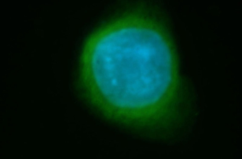
Immunofluorescent analysis of SH-SY5Y cells, using CDKN2A antibody Catalog No:113538 at 1:50 dilution and FITC-labeled donkey anti-rabbit IgG(green). Blue pseudocolor = DAPI (fluorescent DNA dye).
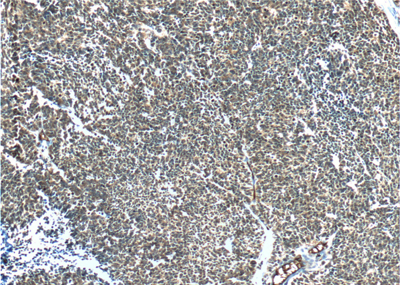
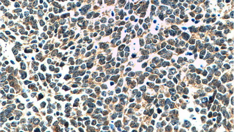
Immunohistochemistry of paraffin-embedded human lung cancer tissue slide using Catalog No:113538(P16-INK4A Antibody) at dilution of 1:200 (under 10x lens).
Immunohistochemistry of paraffin-embedded human lung cancer tissue slide using Catalog No:113538(P16-INK4A Antibody) at dilution of 1:200 (under 40x lens).
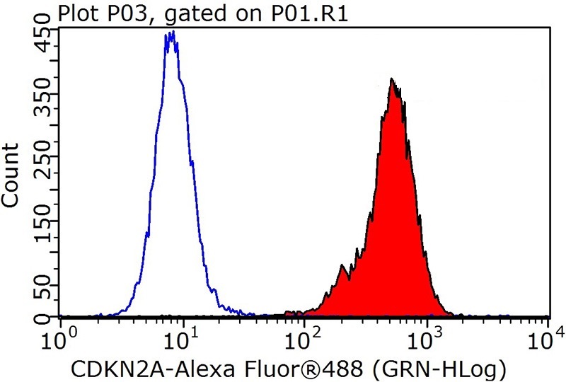
1X10^6 HeLa cells were stained with .2ug P16-INK4A antibody (Catalog No:113538, red) and control antibody (blue). Fixed with 90% MeOH blocked with 3% BSA (30 min). Alexa Fluor 488-congugated AffiniPure Goat Anti-Rabbit IgG(H+L) with dilution 1:1000.
-
Background
CDKN2A generates several transcript variants which differ in their first exons. At least three alternatively-spliced variants encoding distinct proteins have been reported, and two are related, named p16-INK4 and p14 respetively, sharing 50% identity. The third one is completely stuctually un-related, called p14(ARF) or p19(ARF). 10883-1-AP reacted with p16 specifically. P16 plays an important role in regulating the cell cycle, and mutations in p16 increase the risk of developing a variety of cancers, notably melanoma.
-
References
- Motterle A, Pu X, Wood H. Functional analyses of coronary artery disease associated variation on chromosome 9p21 in vascular smooth muscle cells. Human molecular genetics. 21(18):4021-9. 2012.
- Wang F, Xia X, Wang J, Sun Q, Luo J, Cheng B. Notch1 signaling contributes to the oncogenic effect of HBx on human hepatic cells. Biotechnology letters. 35(1):29-37. 2013.
- Xu X, Hueckstaedt LK, Ren J. Deficiency of insulin-like growth factor 1 attenuates aging-induced changes in hepatic function: role of autophagy. Journal of hepatology. 59(2):308-17. 2013.
- Deng S, Hu B, An HM. Teng-Long-Bu-Zhong-Tang, a Chinese herbal formula, enhances anticancer effects of 5--Fluorouracil in CT26 colon carcinoma. BMC complementary and alternative medicine. 13:128. 2013.
- Chidlow G, Wood JP, Sharma S. Ocular expression and distribution of products of the POAG-associated chromosome 9p21 gene region. PloS one. 8(9):e75067. 2013.
- Liang PI, Li CF, Chen LT. BCL6 overexpression is associated with decreased p19 ARF expression and confers an independent prognosticator in gallbladder carcinoma. Tumour biology : the journal of the International Society for Oncodevelopmental Biology and Medicine. 35(2):1417-26. 2014.
- Zhuge CC, Xu JY, Zhang J. Fullerenol protects retinal pigment epithelial cells from oxidative stress-induced premature senescence via activating SIRT1. Investigative ophthalmology & visual science. 55(7):4628-38. 2014.
- Damsky W, Micevic G, Meeth K. mTORC1 activation blocks BrafV600E-induced growth arrest but is insufficient for melanoma formation. Cancer cell. 27(1):41-56. 2015.
Related Products / Services
Please note: All products are "FOR RESEARCH USE ONLY AND ARE NOT INTENDED FOR DIAGNOSTIC OR THERAPEUTIC USE"