
-
Product Name
Lamin B1 antibody
- Documents
-
Description
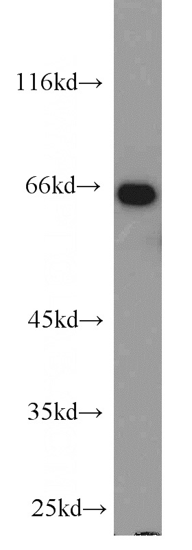
Lamin B1 Rabbit Polyclonal antibody. Positive WB detected in HeLa cells, HepG2 cells, human brain tissue, human kidney tissue, Jurkat cells, K-562 cells, mouse brain tissue, mouse eye tissue, mouse kidney tissue, mouse lung tissue, mouse pancreas tissue, mouse spleen tissue, NIH/3T3 cells, rat brain tissue, rat heart tissue, SH-SY5Y cells, U-937 cells, Y79 cells. Positive IP detected in HeLa cells. Positive FC detected in HEK-293 cells. Positive IHC detected in human breast cancer tissue, human kidney tissue. Positive IF detected in HepG2 cells. Observed molecular weight by Western-blot: 66 kDa
-
Tested applications
ELISA, IHC, IF, IP, WB, FC
-
Species reactivity
Human,Mouse,Rat; other species not tested.
-
Alternative names
ADLD antibody; lamin B1 antibody; LMN antibody; LMN2 antibody; LMNB antibody; LMNB1 antibody
-
Isotype
Rabbit IgG
-
Preparation
This antibody was obtained by immunization of Lamin B1 recombinant protein (Accession Number: NM_005573). Purification method: Antigen affinity purified.
-
Clonality
Polyclonal
-
Formulation
PBS with 0.1% sodium azide and 50% glycerol pH 7.3.
-
Storage instructions
Store at -20℃. DO NOT ALIQUOT
-
Applications
Recommended Dilution:
WB: 1:500-1:5000
IP: 1:500-1:5000
IHC: 1:20-1:200
IF: 1:20-1:200
-
Validations
HeLa cells were subjected to SDS PAGE followed by western blot with Catalog No:117329(LMNB1 antibody) at dilution of 1:1000
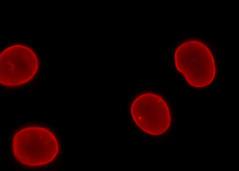
Immunofluorescent analysis of HepG2 cells, using LMNB1 antibody Catalog No:117329 at 1:50 dilution and Rhodamine-labeled goat anti-rabbit IgG (red).
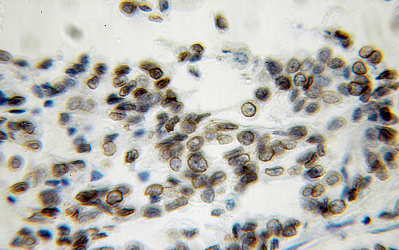
Immunohistochemical of paraffin-embedded human breast cancer using Catalog No:117329(LMNB1 antibody) at dilution of 1:100 (under 10x lens)
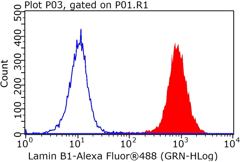
1X10^6 HEK-293 cells were stained with 0.2ug Lamin B1 antibody (Catalog No:117329, red) and control antibody (blue). Fixed with 90% MeOH blocked with 3% BSA (30 min). FITC-Goat anti-Rabbit IgG with dilution 1:100.
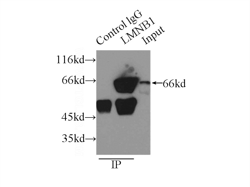
IP Result of anti-LMNB1 (IP:Catalog No:117329, 3ug; Detection:Catalog No:117329 1:1000) with HeLa cells lysate 2500ug.
-
Background
Lamins are components of the nuclear lamina, a fibrous layer on the nucleoplasmic side of the inner nuclear membrane, which is thought to provide a framework for the nuclear envelope and may also interact with chromatin. The nuclear lamina consists of a two-dimensional matrix of proteins located next to the inner nuclear membrane. The lamin family of proteins make up the matrix and are highly conserved in evolution. During mitosis, the lamina matrix is reversibly disassembled as the lamin proteins are phosphorylated. Vertebrate lamins consist of two types, A and B. This gene encodes one of the two B type proteins, B1. Expression of uncleavable mutant lamin A or B caused significant delays in the onset of chromatin condensation and nuclear shrinkage during apoptosis(PMID:11953316). This protein is not suitable for samples where the nuclear envelope has been removed.
-
References
- Yeh JL, Hsu JH, Wu PJ. KMUP-1 attenuates isoprenaline-induced cardiac hypertrophy in rats through NO/cGMP/PKG and ERK1/2/calcineurin A pathways. British journal of pharmacology. 159(5):1151-60. 2010.
- Chen Y, Guan Y, Liu H. Activation of the Wnt/β-catenin signaling pathway is associated with glial proliferation in the adult spinal cord of ALS transgenic mice. Biochemical and biophysical research communications. 420(2):397-403. 2012.
- Chen H, Li H, Cao F. 1,2,3,4,6-penta-O-galloyl-β-D-glucose protects PC12 Cells from MPP(+)-mediated cell death by inducing heme oxygenase-1 in an ERK- and Akt-dependent manner. Journal of Huazhong University of Science and Technology. Medical sciences = Hua zhong ke ji da xue xue bao. Yi xue Ying De wen ban = Huazhong keji daxue xuebao. Yixue Yingdewen ban. 32(5):737-45. 2012.
- Li Y, Chen R, Zhou Q. LSm14A is a processing body-associated sensor of viral nucleic acids that initiates cellular antiviral response in the early phase of viral infection. Proceedings of the National Academy of Sciences of the United States of America. 109(29):11770-5. 2012.
- Shi H, Dong L, Jiang J. Chlorogenic acid reduces liver inflammation and fibrosis through inhibition of toll-like receptor 4 signaling pathway. Toxicology. 303:107-14. 2013.
- Wang J, Li W, Wang R, Xue J, Chen J. Adeno-associated virus Rep78 restricts adenovirus E1B55K-mediated p53 nuclear exportation. Acta biochimica et biophysica Sinica. 45(2):135-40. 2013.
- Shan Y, Schoenfeld RA, Hayashi G. Frataxin deficiency leads to defects in expression of antioxidants and Nrf2 expression in dorsal root ganglia of the Friedreich's ataxia YG8R mouse model. Antioxidants & redox signaling. 19(13):1481-93. 2013.
- Tu JB, Dong Q, Hu XY. Proteomic analysis of mitochondria from infantile hemangioma endothelial cells treated with sodium morrhuate and its liposomal formulation. Journal of biochemical and molecular toxicology. 26(9):374-80. 2012.
Related Products / Services
Please note: All products are "FOR RESEARCH USE ONLY AND ARE NOT INTENDED FOR DIAGNOSTIC OR THERAPEUTIC USE"