
-
Product Name
Lamin A/C antibody
- Documents
-
Description
Lamin A/C Rabbit Polyclonal antibody. Positive FC detected in HEK-293T cells. Positive IF detected in HepG2 cells. Positive IP detected in A375 cells. Positive WB detected in NIH/3T3 cells, A375 cells, C6 cells, HEK-293 cells, HeLa cells, mouse heart tissue, mouse ovary tissue, SKOV-3 cells. Observed molecular weight by Western-blot: 65 kDa,70 kDa
-
Tested applications
ELISA, IP, FC, IF, WB
-
Species reactivity
Human,Mouse,Rat; other species not tested.
-
Alternative names
70 kDa lamin antibody; CDCD1 antibody; CDDC antibody; CMD1A antibody; CMT2B1 antibody; EMD2 antibody; FPL antibody; FPLD antibody; HGPS antibody; IDC antibody; lamin A antibody; lamin A/C antibody; LDP1 antibody; LFP antibody; LGMD1B antibody; LMN1 antibody; LMNA antibody; LMNC antibody; Prelamin A/C antibody; PRO1 antibody
-
Isotype
Rabbit IgG
-
Preparation
This antibody was obtained by immunization of Lamin A/C recombinant protein (Accession Number: NM_001406992). Purification method: Antigen affinity purified.
-
Clonality
Polyclonal
-
Formulation
PBS with 0.1% sodium azide and 50% glycerol pH 7.3.
-
Storage instructions
Store at -20℃. DO NOT ALIQUOT
-
Applications
Recommended Dilution:
WB: 1:1000-1:10000
IP: 1:500-1:5000
IF: 1:20-1:200
-
Validations
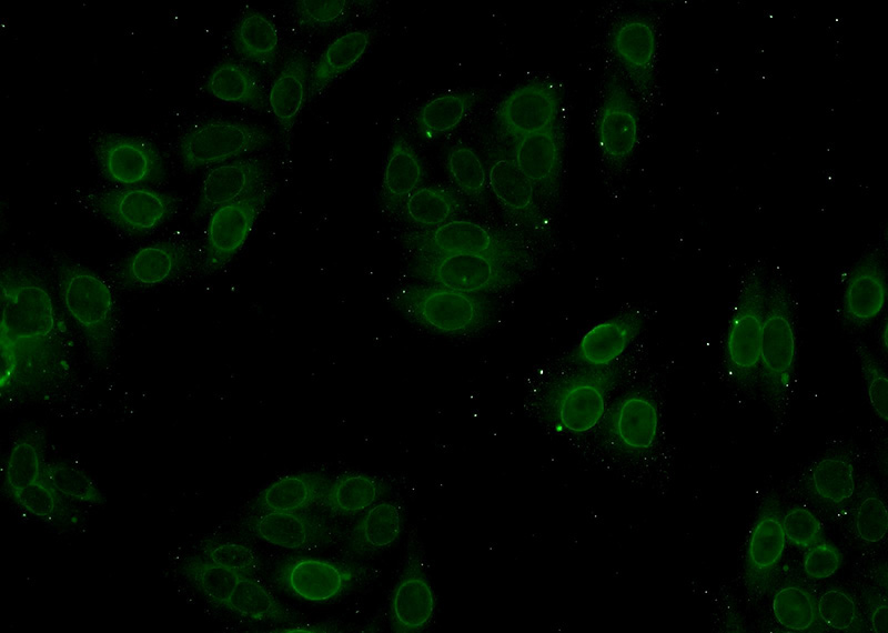
Immunofluorescent analysis of HepG2 cells using Catalog No:117327(Lamin A/C Antibody) at dilution of 1:50 and Alexa Fluor 488-congugated AffiniPure Goat Anti-Rabbit IgG(H+L)
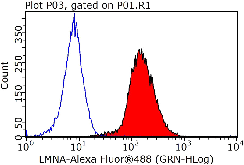
1X10^6 HEK-293T cells were stained with 0.2ug Lamin A/C antibody (Catalog No:117327, red) and control antibody (blue). Fixed with 90% MeOH blocked with 3% BSA (30 min). Alexa Fluor 488-congugated AffiniPure Goat Anti-Rabbit IgG(H+L) with dilution 1:1000.
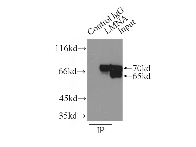
IP Result of anti-lamin-A (IP:Catalog No:117327, 3ug; Detection:Catalog No:117327 1:1000) with A375 cells lysate 800ug.
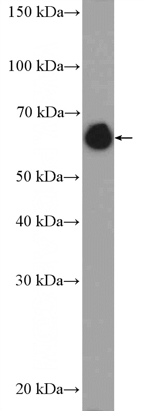
NIH/3T3 cells were subjected to SDS PAGE followed by western blot with Catalog No:117327(Lamin A/C Antibody) at dilution of 1:3000
-
Background
Lamin A/C is also named as LMNA, FPL, LFP, EMD2, FPLD, HGPS, LDP1, LMN1. The lamin family of proteins make up the matrix and are highly conserved in evolution. During mitosis, the lamina matrix is reversibly disassembled as the lamin proteins are phosphorylated. Lamin proteins are thought to be involved in nuclear stability, chromatin structure and gene expression. The lack of lamin A/C can be as a novel marker for undifferentiated embryonic stem cells and lamin A/C expression is as an early indicator of differentiation(PMID: 16179429). Mutations in this gene lead to several diseases: Emery-Dreifuss muscular dystrophy, familial partial lipodystrophy, limb girdle muscular dystrophy, dilated cardiomyopathy, Charcot-Marie-Tooth disease, and Hutchinson-Gilford progeria syndrome. This protein has 4 isoforms produced by alternative splicing with the molecular weight of 74 kDa, 65 kDa, 70 kDa and 64 kDa. This antibody can recognize 4 isoforms of Lamin A/C.
-
References
- Nakamura M, Morisawa H, Imajoh-Ohmi S, Takamura C, Fukuda H, Toda T. Proteomic analysis of protein complexes in human SH-SY5Y neuroblastoma cells by using blue-native gel electrophoresis: an increase in lamin A/C associated with heat shock protein 90 in response to 6-hydroxydopamine-induced oxidative stress. Experimental gerontology. 44(6-7):375-82. 2009.
- Zhao Y, Ben H, Qu S. Proteomic analysis of primary duck hepatocytes infected with duck hepatitis B virus. Proteome science. 8:28. 2010.
- Sun X, Lu B, Hu B, Xiao W, Li W, Huang Z. Novel function of the chromosome 7 open reading frame 41 gene to promote leukemic megakaryocyte differentiation by modulating TPA-induced signaling. Blood cancer journal. 4:e198. 2014.
- Shen M, Chen K, Lu J. Protective effect of astaxanthin on liver fibrosis through modulation of TGF-β1 expression and autophagy. Mediators of inflammation. 2014:954502. 2014.
- Lu Y, Cai G, Cui S. FHL2-driven molecular network mediated Septin2 knockdown inducing apoptosis in mesangial cell. Proteomics. 14(21-22):2485-97. 2014.
- Lu B, Sun X, Chen Y. Novel function of PITH domain-containing 1 as an activator of internal ribosomal entry site to enhance RUNX1 expression and promote megakaryocyte differentiation. Cellular and molecular life sciences : CMLS. 72(4):821-32. 2015.
- Duan X, Zohaib A, Li Y. miR-206 modulates lipopolysaccharide-mediated inflammatory cytokine production in human astrocytes. Cellular signalling. 27(1):61-8. 2015.
- Wen Y, Li J, Wang L. UDP-glucose dehydrogenase modulates proteoglycan synthesis in articular chondrocytes: its possible involvement and regulation in osteoarthritis. Arthritis research & therapy. 16(6):484. 2014.
Related Products / Services
Please note: All products are "FOR RESEARCH USE ONLY AND ARE NOT INTENDED FOR DIAGNOSTIC OR THERAPEUTIC USE"