-
Product Name
JNK antibody
- Documents
-
Description
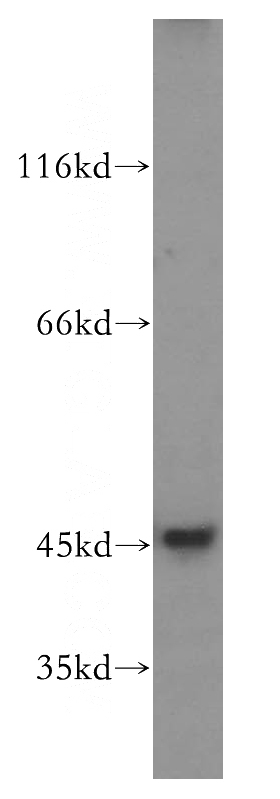
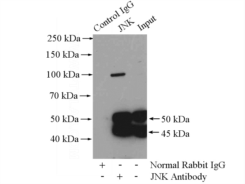
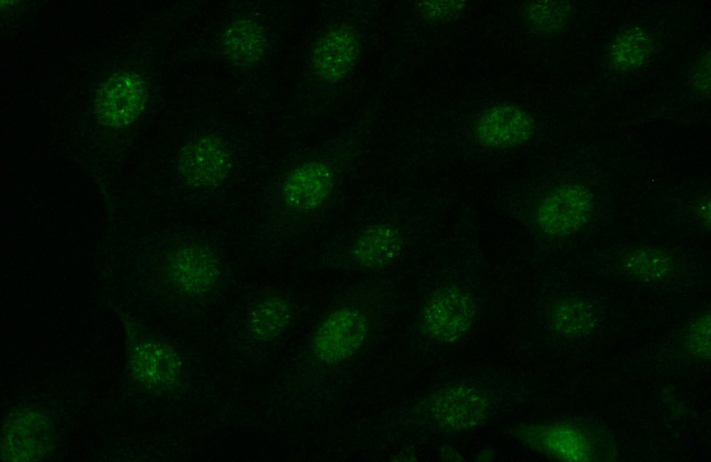
JNK Rabbit Polyclonal antibody. Positive IF detected in SH-SY5Y cells. Positive IP detected in HEK-293 cells. Positive WB detected in SH-SY5Y cells, A431 cells, HEK-293 cells, HeLa cells, HT-1080 cells, PC-3 cells. Observed molecular weight by Western-blot: 44-48 kDa
-
Tested applications
ELISA, WB, IP, IF
-
Species reactivity
Human, Mouse; other species not tested.
-
Alternative names
c Jun N terminal kinase 1 antibody; JNK antibody; JNK 46 antibody; JNK1 antibody; JNK1A2 antibody; JNK21B1/2 antibody; MAP kinase 8 antibody; MAPK 8 antibody; MAPK8 antibody; PRKM8 antibody; SAPK1 antibody
-
Isotype
Rabbit IgG
-
Preparation
This antibody was obtained by immunization of Peptide (Accession Number: NM_001323323). Purification method: Antigen affinity purified.
-
Clonality
Polyclonal
-
Formulation
PBS with 0.02% sodium azide and 50% glycerol pH 7.3.
-
Storage instructions
Store at -20℃. DO NOT ALIQUOT
-
Applications
Recommended Dilution:
WB: 1:500-1:5000
IP: 1:200-1:2000
IF: 1:50-1:500
-
Validations

SH-SY5Y cells were subjected to SDS PAGE followed by western blot with Catalog No:111892(JNK antibody) at dilution of 1:300

IP Result of anti-JNK (IP:Catalog No:111892, 4ug; Detection:Catalog No:111892 1:600) with HEK-293 cells lysate 2000ug.

Immunofluorescent analysis of (-20oc Acetone) fixed SH-SY5Y cells using Catalog No:111892(JNK Antibody) at dilution of 1:50 and Alexa Fluor 488-congugated AffiniPure Goat Anti-Rabbit IgG(H+L)
-
Background
MAPK8(Mitogen-activated protein kinase 8) is also named as JNK1, PRKM8, SAPK1, SAPK1C and belongs to the MAP kinase subfamily. MAPK8 is activated by dual phosphorylation at a Thr-Pro-Tyr motif during response to UV light. MAPK8 functions to phosphorylate c-Jun at N-terminal serine regulatory sites of Ser-63 and Ser-73, mapping within the transactivation domain. Phosphorylation of these sites in response to UV results in transcriptional activation of c-Jun. It has 4 isoforms produced by alternative splicing with the molecular weight of 46 kDa and 48 kDa. This protein can be phosphorylated and this antibody recognizes the 46 kDa and 55 kDa bands in western blot(PMID:11062067 ).
-
References
- Wu J, Huang Z, Ren J. Mlkl knockout mice demonstrate the indispensable role of Mlkl in necroptosis. Cell research. 23(8):994-1006. 2013.
- Sun L, Liu S, Sun Q. Inhibition of TROY promotes OPC differentiation and increases therapeutic efficacy of OPC graft for spinal cord injury. Stem cells and development. 23(17):2104-18. 2014.
- Yang CC, Yao CA, Yang JC, Chien CT. Sialic acid rescues repurified lipopolysaccharide-induced acute renal failure via inhibiting TLR4/PKC/gp91-mediated endoplasmic reticulum stress, apoptosis, autophagy, and pyroptosis signaling. Toxicological sciences : an official journal of the Society of Toxicology. 141(1):155-65. 2014.
- Xu D, Su C, Song X. Polychlorinated biphenyl quinone induces endoplasmic reticulum stress, unfolded protein response, and calcium release. Chemical research in toxicology. 28(6):1326-37. 2015.
- Su C, Xia X, Shi Q. Neohesperidin Dihydrochalcone versus CCl₄-Induced Hepatic Injury through Different Mechanisms: The Implication of Free Radical Scavenging and Nrf2 Activation. Journal of agricultural and food chemistry. 63(22):5468-75. 2015.
- Li J, Wang F, Xia Y. Astaxanthin Pretreatment Attenuates Hepatic Ischemia Reperfusion-Induced Apoptosis and Autophagy via the ROS/MAPK Pathway in Mice. Marine drugs. 13(6):3368-87. 2015.
- Li Q, Fan YS, Gao ZQ, Fan K, Liu ZJ. Effect of Fructus Ligustri Lucidi on osteoblastic like cell-line MC3T3-E1. Journal of ethnopharmacology. 170:88-95. 2015.
- Gu A, Jie Y, Sun L, Zhao S, E M, You Q. RhNRG-1β Protects the Myocardium against Irradiation-Induced Damage via the ErbB2-ERK-SIRT1 Signaling Pathway. PloS one. 10(9):e0137337. 2015.
Related Products / Services
Please note: All products are "FOR RESEARCH USE ONLY AND ARE NOT INTENDED FOR DIAGNOSTIC OR THERAPEUTIC USE"
