-
Product Name
GLAST antibody
- Documents
-
Description
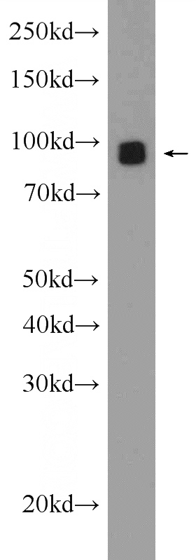
GLAST Rabbit Polyclonal antibody. Positive WB detected in Neuro-2a cells, C6 cells, mouse brain tissue. Positive IHC detected in human brain tissue, mouse brain tissue. Observed molecular weight by Western-blot: 50-55, 90-100 kDa
-
Tested applications
ELISA, IHC, WB
-
Species reactivity
Human, Mouse, Rat; other species not tested.
-
Alternative names
EA6 antibody; EAAT1 antibody; GLAST antibody; GLAST 1 antibody; GLAST1 antibody; SLC1A3 antibody
- Immunogen
-
Isotype
Rabbit IgG
-
Preparation
This antibody was obtained by immunization of GLAST recombinant protein (Accession Number: NM_004172). Purification method: Antigen affinity purified.
-
Clonality
Polyclonal
-
Formulation
PBS with 0.1% sodium azide and 50% glycerol pH 7.3.
-
Storage instructions
Store at -20℃. DO NOT ALIQUOT
-
Applications
Recommended Dilution:
WB: 1:500-1:5000
IHC: 1:20-1:200
-
Validations

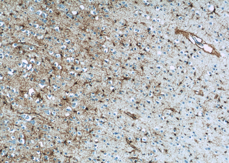
Immunohistochemistry of paraffin-embedded human brain tissue slide using Catalog No:111019(SLC1A3 Antibody) at dilution of 1:50 (under 10x lens)

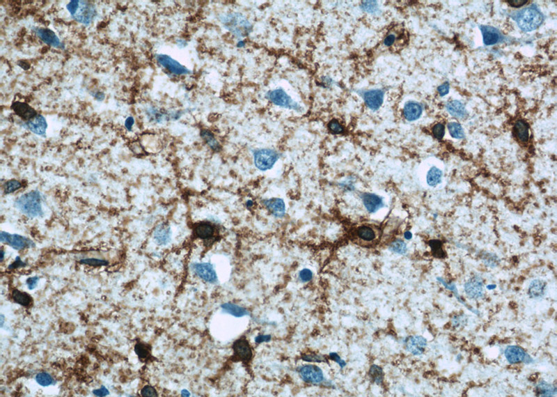
Immunohistochemistry of paraffin-embedded human brain tissue slide using Catalog No:111019(SLC1A3 Antibody) at dilution of 1:50 (under 40x lens)

Neuro-2a cells were subjected to SDS PAGE followed by western blot with Catalog No:111019(SLC1A3 Antibody) at dilution of 1:1000
-
Background
SLC1A3, also known as EAAT-1 or GLAST, is a membrane-bound protein localized in glial cells and pre-synaptic glutamatergic nerve endings. It transports the excitatory neurotransmitters L-glutamate and D-aspartate, which is essential for terminating the postsynaptic acction of glutamate. Recently, a correlation between expression/function of glial EAAT-1 and tumor proliferation has been reported. The exceptionally rare expression of EAAT-1 in non-neoplastic choroid plexus (CP) compared to choroid plexus tumors (CPT) may distinguishes neoplastic from normal CP. There are a number of splicing variants of SLC1A3, like GLAST1a and GLAST1b, exist due to the exon skipping. It also undergo glycosylation. Variety of bands can be observed in the western blotting assay: 50-55 kDa represents the unglycosylated GLAST1a or GLAST1b, 65-70 kDa correspond to the glycosylated proteins, larger proteins between 90-130 kDa may be the multimers of SLC1A3. (11086157, 17471058, 12546822)
Related Products / Services
Please note: All products are "FOR RESEARCH USE ONLY AND ARE NOT INTENDED FOR DIAGNOSTIC OR THERAPEUTIC USE"
