
-
Product Name
FBXW7 Polyclonal Antibody
- Documents
-
Description
Polyclonal antibody to FBXW7
-
Tested applications
WB, IHC, IF
-
Species reactivity
Human, Mouse, Rat
-
Alternative names
FBXW7 antibody; AGO antibody; CDC4 antibody; FBW6 antibody; FBW7 antibody; FBX30 antibody; FBXO30 antibody; FBXW6 antibody; SEL-10 antibody; SEL10 antibody; hAgo antibody; hCdc4 antibody; F-box/WD repeat-containing protein 7 antibody
-
Isotype
Rabbit IgG
-
Preparation
Antigen: Recombinant fusion protein containing a sequence corresponding to amino acids 408-707 of human FBXW7 (NP_361014.1).
-
Clonality
Polyclonal
-
Formulation
PBS with 0.02% sodium azide, 50% glycerol, pH7.3.
-
Storage instructions
Store at -20℃. Avoid freeze / thaw cycles.
-
Applications
WB 1:500 - 1:2000
IHC 1:50 - 1:200
IF 1:50 - 1:200 -
Validations
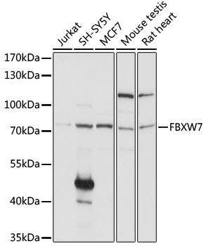
Western blot - FBXW7 Polyclonal Antibody
Western blot analysis of extracts of various cell lines, using FBXW7 antibody at 1:1000 dilution.Secondary antibody: HRP Goat Anti-Rabbit IgG (H+L) at 1:10000 dilution.Lysates/proteins: 25ug per lane.Blocking buffer: 3% nonfat dry milk in TBST.Detection: ECL Basic Kit .Exposure time: 60s.
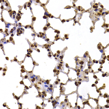
Immunohistochemistry - FBXW7 Polyclonal Antibody
Immunohistochemistry of paraffin-embedded mouse lung using FBXW7 antibody at dilution of 1:100 (40x lens).
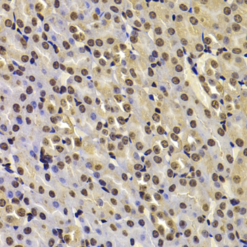
Immunohistochemistry - FBXW7 Polyclonal Antibody
Immunohistochemistry of paraffin-embedded mouse kidney using FBXW7 antibody at dilution of 1:100 (40x lens).
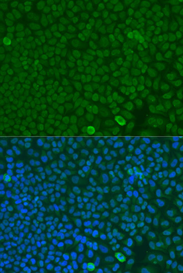
Immunofluorescence - FBXW7 Polyclonal Antibody
Immunofluorescence analysis of U2OS cells using FBXW7 antibody at dilution of 1:100. Blue: DAPI for nuclear staining.
-
Background
Substrate recognition component of a SCF (SKP1-CUL1-F-box protein) E3 ubiquitin-protein ligase complex which mediates the ubiquitination and subsequent proteasomal degradation of target proteins. Recognizes and binds phosphorylated sites/phosphodegrons within target proteins and thereafter bring them to the SCF complex for ubiquitination. Identified substrates include cyclin-E (CCNE1 or CCNE2), DISC1, JUN, MYC, NOTCH1 released notch intracellular domain (NICD), NOTCH2, MCL1, and probably PSEN1. Acts as a negative regulator of JNK signaling by binding to phosphorylated JUN and promoting its ubiquitination and subsequent degradation. SCF(FBXW7) complex mediates the ubiquitination and subsequent degradation of NFE2L1 (By similarity). Involved in bone homeostasis and negative regulation of osteoclast differentiation.
Related Products / Services
Please note: All products are "FOR RESEARCH USE ONLY AND ARE NOT INTENDED FOR DIAGNOSTIC OR THERAPEUTIC USE"