-
Product Name
CFHR3 antibody
- Documents
-
Description
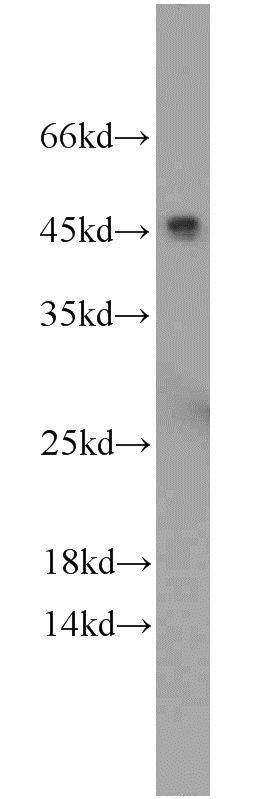
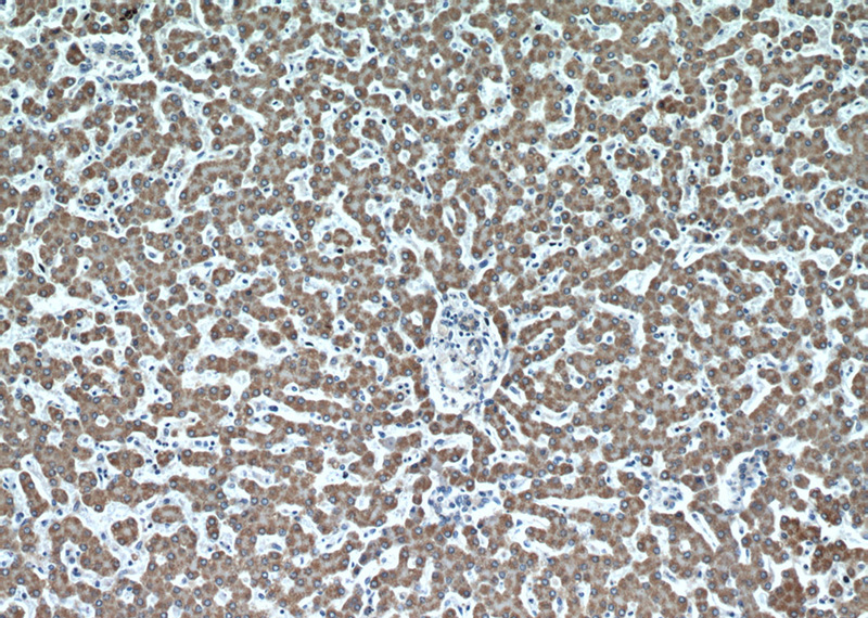
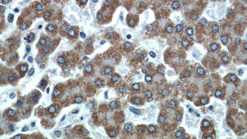
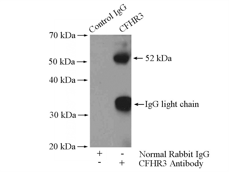
CFHR3 Rabbit Polyclonal antibody. Positive IHC detected in human liver tissue. Positive WB detected in human heart tissue. Positive IP detected in HepG2 cells. Observed molecular weight by Western-blot: 45-56 kDa
-
Tested applications
ELISA, WB, IP, IHC
-
Species reactivity
Human; other species not tested.
-
Alternative names
CFHL3 antibody; CFHR3 antibody; complement factor H related 3 antibody; DOWN16 antibody; FHR 3 antibody; FHR3 antibody; H factor like protein 3 antibody; HLF4 antibody
-
Isotype
Rabbit IgG
-
Preparation
This antibody was obtained by immunization of CFHR3 recombinant protein (Accession Number: NM_021023). Purification method: Antigen affinity purified.
-
Clonality
Polyclonal
-
Formulation
PBS with 0.02% sodium azide and 50% glycerol pH 7.3.
-
Storage instructions
Store at -20℃. DO NOT ALIQUOT
-
Applications
Recommended Dilution:
WB: 1:500-1:5000
IP: 1:200-1:1000
IHC: 1:50-1:500
-
Validations

human heart tissue were subjected to SDS PAGE followed by western blot with Catalog No:109198(CFHR3 antibody) at dilution of 1:2000

Immunohistochemical of paraffin-embedded human liver using Catalog No:109198(CFHR3 antibody) at dilution of 1:50 (under 10x lens)

Immunohistochemical of paraffin-embedded human liver using Catalog No:109198(CFHR3 antibody) at dilution of 1:50 (under 40x lens)

IP Result of anti-CFHR3 (IP:Catalog No:109198, 4ug; Detection:Catalog No:109198 1:300) with HepG2 cells lysate 2400ug.
-
Background
CFHR3, also named as DOWN16, belongs to the complement factor H-related protein family. Expressed by the liver and secreted in plasma, human CFHR3 is composed of five short consensus repeats (SCRs), and it also has a 19-amino acid signal peptide and four N-linked glycosylation sites. It may be involved in complement regualtion. A frequent deletion of CFHR1 and CFHR3 genes was found to be associated with decrease risk of aged-related macular degeneration (AMD), and with an increased risk of atypical hemolytic-uremic syndrome (aHUS).
-
References
- Yoshida Y, Miyata T, Matsumoto M. A novel quantitative hemolytic assay coupled with restriction fragment length polymorphisms analysis enabled early diagnosis of atypical hemolytic uremic syndrome and identified unique predisposing mutations in Japan. PloS one. 10(5):e0124655. 2015.
Related Products / Services
Please note: All products are "FOR RESEARCH USE ONLY AND ARE NOT INTENDED FOR DIAGNOSTIC OR THERAPEUTIC USE"
