
-
Product Name
ATP5H antibody
- Documents
-
Description
ATP5H Rabbit Polyclonal antibody. Positive IHC detected in human lung cancer tissue, human pancreas tissue. Positive IF detected in HepG2 cells. Positive IP detected in mouse liver tissue. Positive WB detected in Jurkat cells, human brain tissue, human liver tissue, mouse brain tissue, mouse liver tissue, mouse ovary tissue, rat brain tissue, rat liver tissue. Observed molecular weight by Western-blot: 19-22 kDa
-
Tested applications
ELISA, IHC, IF, WB, IP
-
Species reactivity
Human, Mouse, Rat; other species not tested.
-
Alternative names
ATP5H antibody; ATP5JD antibody; ATPase subunit d antibody; ATPQ antibody
-
Isotype
Rabbit IgG
-
Preparation
This antibody was obtained by immunization of ATP5H recombinant protein (Accession Number: NM_001003785). Purification method: Antigen affinity purified.
-
Clonality
Polyclonal
-
Formulation
PBS with 0.02% sodium azide and 50% glycerol pH 7.3.
-
Storage instructions
Store at -20℃. DO NOT ALIQUOT
-
Applications
Recommended Dilution:
WB: 1:1000-1:10000
IP: 1:1000-1:10000
IHC: 1:20-1:200
IF: 1:10-1:100
-
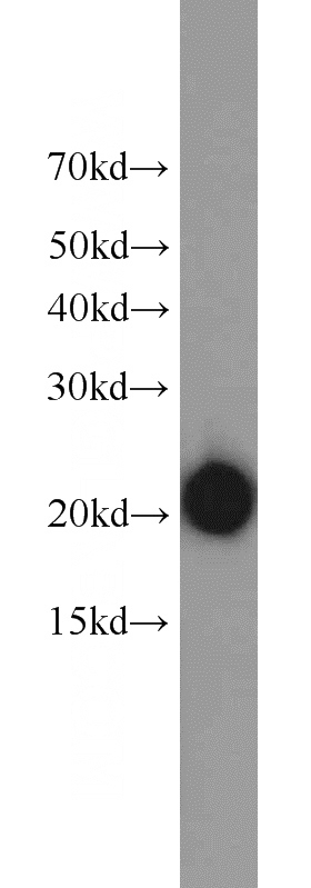
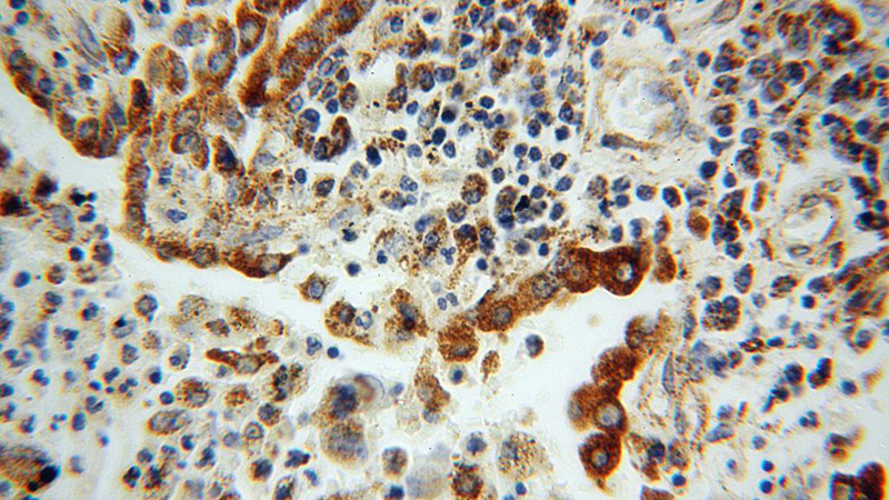
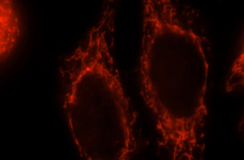
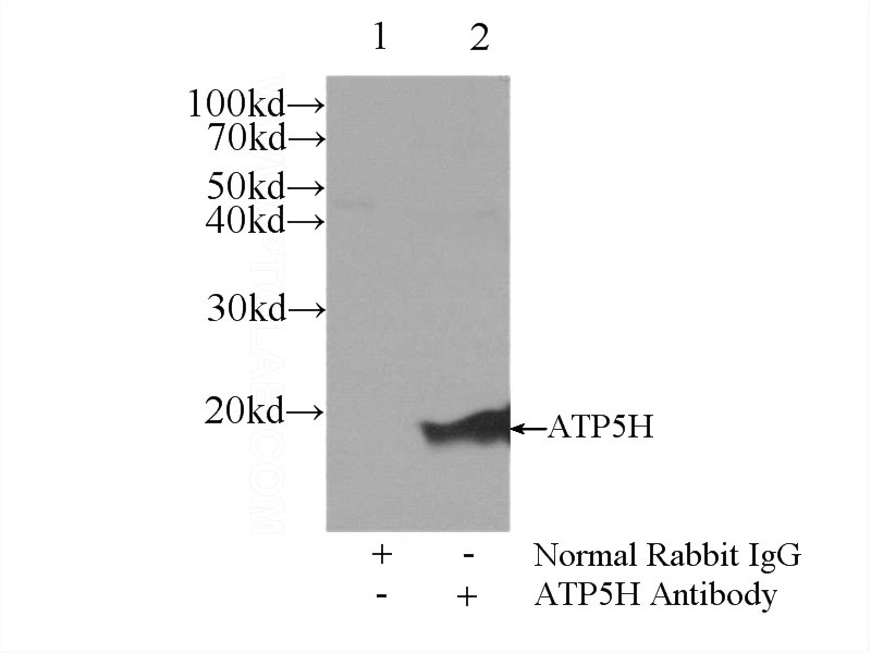
Validations
Jurkat cells were subjected to SDS PAGE followed by western blot with Catalog No:108355(ATP5H antibody) at dilution of 1:3000
Immunohistochemical of paraffin-embedded human lung cancer using Catalog No:108355(ATP5H antibody) at dilution of 1:100 (under 40x lens)
Immunofluorescent analysis of HepG2 cells, using ATP5H antibody Catalog No:108355 at 1:25 dilution and Rhodamine-labeled goat anti-rabbit IgG (red).
IP Result of anti-ATP5H (IP:Catalog No:108355, 3ug; Detection:Catalog No:108355 1:2000) with mouse liver tissue lysate 6000ug.
-
Background
Mitochondrial membrane ATP synthase (F1-Fo ATP synthase or Complex V) produces ATP from ADP in the presence of a proton gradient across the membrane which is generated by electron transport complexes of the respiratory chain. It is composed of the soluble catalytic core, F1, and the membrane-spanning component and Fo, which comprises the proton channel. The Fo seems to have nine subunits (a, b, c, d, e, f, g, F6 and 8). ATP5H gene encodes ATP synthase subunit d of the Fo complex.
Related Products / Services
Please note: All products are "FOR RESEARCH USE ONLY AND ARE NOT INTENDED FOR DIAGNOSTIC OR THERAPEUTIC USE"