
-
Product Name
Anti-YBX2 antibody
- Documents
-
Description
Rabbit polyclonal antibody to YBX2
-
Tested applications
WB, ICC, IHC-P, FC
-
Species reactivity
Human, Mouse, Rat
-
Alternative names
DBPC antibody; MSY2 antibody; CSDA3 antibody; CONTRIN antibody
-
Isotype
Rabbit IgG
-
Preparation
This antigen of this antibody was synthetic peptide within human ybox1 aa 302-355.
-
Clonality
Polyclonal
-
Formulation
Liquid, 1*PBS (pH7.4), 0.2% BSA, 40% Glycerol. Preservative: 0.05% Sodium Azide.
-
Storage instructions
Store at +4℃ after thawing. Aliquot store at -20℃ or -80℃. Avoid repeated freeze / thaw cycles.
-
Applications
WB: 1:1,000
ICC: 1:50-1:200
IHC-P: 1:50-1:200
FC: 1:50-1:100
-
Validations
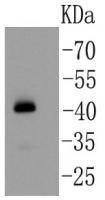
Fig1: Western blot analysis on mouse testis lysates using anti-YB1 rabbit polyclonal antibody.
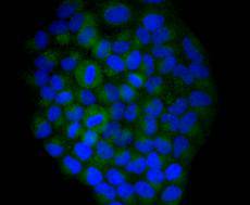
Fig2: Immunocytochemical staining of Hela cells using anti-YB1 rabbit polyclonal antibody.
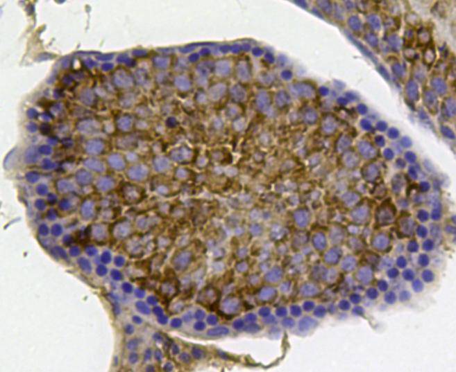
Fig3: Immunohistochemical analysis of paraffin- embedded mouse testis tissue using anti-YB1 rabbit polyclonal antibody.
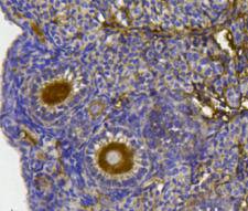
Fig4: Immunohistochemical analysis of paraffin- embedded mouse ovary tissue using anti-YB1 rabbit polyclonal antibody.
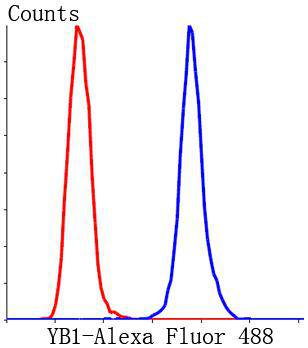
Fig5: Flow cytometric analysis of Hela cells with YB1 antibody at 1/50 dilution (blue) compared with an unlabelled control (cells without incubation with primary antibody; red). Alexa Fluor 488-conjugated Goat anti rabbit IgG was used as the secondary antibody.
- Background
-
References
- Chalmey, C. et al. 2013. Systemic compensatory response to neonatal estradiol exposure does not prevent depletion of the oocyte pool in the rat. PloS one. 8: e82175.
- Shi, K. et al. 2007. Advanced methods of isolation and identification of porcine primordial follicles. Anim Reprod Sci. 101: 163-171.
Related Products / Services
Please note: All products are "FOR RESEARCH USE ONLY AND ARE NOT INTENDED FOR DIAGNOSTIC OR THERAPEUTIC USE"