
-
Product Name
Anti-TUBE1 antibody
- Documents
-
Description
Mouse monoclonal antibody to TUBE1
-
Tested applications
WB, IHC-P, FC
-
Species reactivity
Human
-
Alternative names
TUBE antibody; dJ142L7.2 antibody
-
Isotype
Mouse IgG1
-
Preparation
This antigen of this antibody was recombinant protein
-
Clonality
Monoclonal
-
Formulation
Liquid, 1*TBS (pH7.4), 1%BSA, 40%Glycerol. Preservative: 0.05% Sodium Azide.
-
Storage instructions
Store at +4℃ after thawing. Aliquot store at -20℃ or -80℃. Avoid repeated freeze / thaw cycles.
-
Applications
WB: 1:500-1:2,000
IHC-P: 1:50-1:200
FC: 1:50-1:100
-
Validations
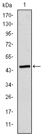
Fig1: Western blot analysis of TUBE1 on human TUBE1 recombinant protein using anti-TUBE1 antibody at 1/1,000 dilution.
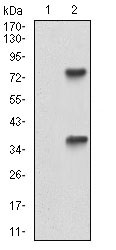
Fig2: Western blot analysis of TUBE1 on HEK293 (1) and TUBE1-hIgGFc transfected HEK293 (2) cell lysate using anti-TUBE1 antibody at 1/1,000 dilution.
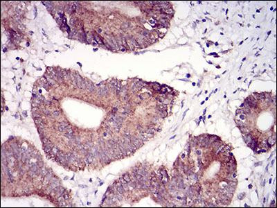
Fig3: Immunohistochemical analysis of paraffin-embedded human colon cancer tissue using anti-TUBE1 antibody. Counter stained with hematoxylin.
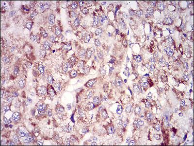
Fig4: Immunohistochemical analysis of paraffin-embedded human liver cancer tissue using anti-TUBE1 antibody. Counter stained with hematoxylin.
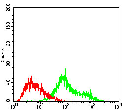
Fig5: Flow cytometric analysis of Hela cells with TUBE1 antibody at 1/100 dilution (green) compared with an unlabelled control (cells without incubation with primary antibody; red).
- Background
-
References
- Chang P et al. Epsilon-tubulin is required for centriole duplication and microtubule organization. Nat Cell Biol. 2003 Jan;5(1):71-6.
- Chang P et al. Delta-tubulin and epsilon-tubulin: two new human centrosomal tubulins reveal new aspects of centrosome structure and function. Nat Cell Biol. 2000 Jan;2(1):30-5.
Related Products / Services
Please note: All products are "FOR RESEARCH USE ONLY AND ARE NOT INTENDED FOR DIAGNOSTIC OR THERAPEUTIC USE"