
-
Product Name
Anti-TESPA1 antibody
- Documents
-
Description
Rabbit polyclonal antibody to TESPA1
-
Tested applications
WB, ICC, IHC-P, FC
-
Species reactivity
Human, Mouse, Rat
-
Alternative names
HSPC257 antibody; ITPRID3 antibody; KIAA0748 antibody
-
Isotype
Rabbit IgG
-
Preparation
This antigen of this antibody was synthetic peptide within human tespa1 aa 226-270.
-
Clonality
Polyclonal
-
Formulation
Liquid, 1*PBS (pH7.4), 0.2% BSA, 40% Glycerol. Preservative: 0.05% Sodium Azide.
-
Storage instructions
Store at +4℃ after thawing. Aliquot store at -20℃. Avoid repeated freeze / thaw cycles.
-
Applications
WB: 1:500-1:1,000
ICC: 1:200
IHC-P: 1:50
FC: 1:50-1:100
-
Validations
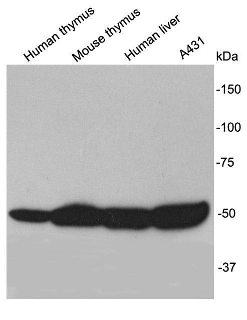
Fig1: Western blot analysis of TESPA1 on different lysates using anti-TESPA1 antibody at 1/1,000 dilution.
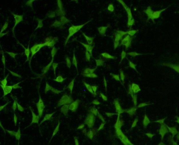
Fig2: ICC staining TESPA1 in A172 cells (green). The nuclear counter stain is DAPI (blue). Cells were fixed in paraformaldehyde, permeabilised with 0.25% Triton X100/PBS.
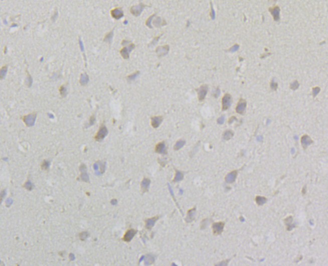
Fig3: Immunohistochemical analysis of paraffin-embedded rat brain tissue using anti-TESPA1 antibody. Counter stained with hematoxylin.
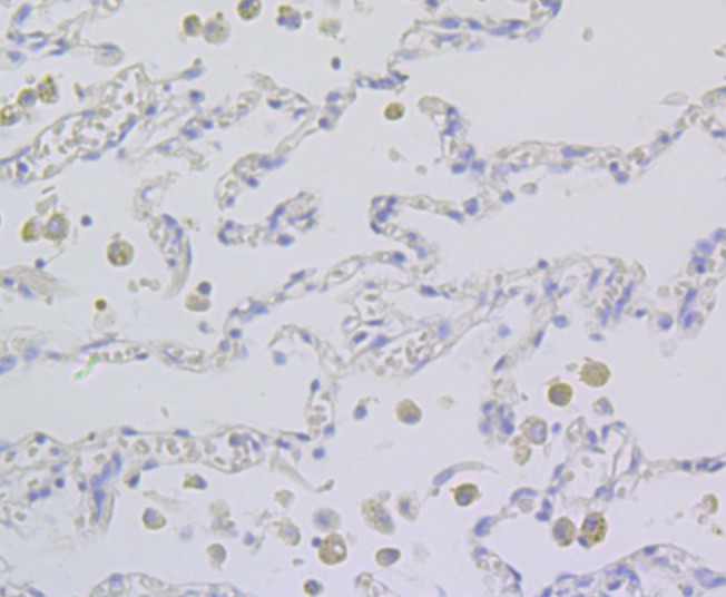
Fig4: Immunohistochemical analysis of paraffin-embedded human lung cancer tissue using anti-TESPA1 antibody. Counter stained with hematoxylin.
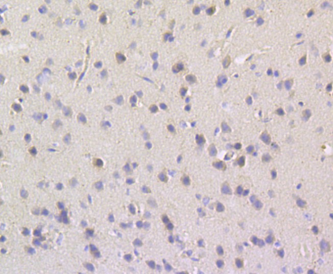
Fig5: Immunohistochemical analysis of paraffin-embedded mouse brain tissue using anti-TESPA1 antibody. Counter stained with hematoxylin.
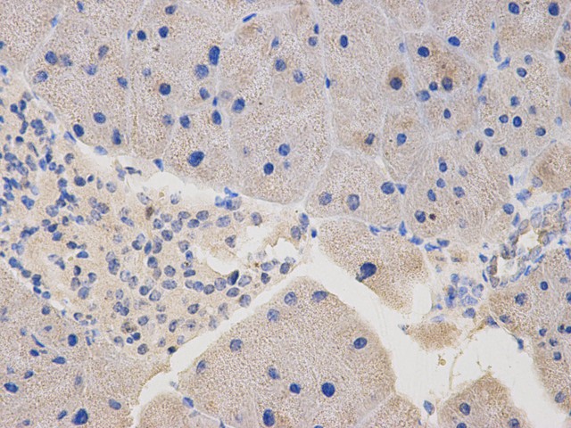
Fig6: Immunohistochemical analysis of paraffin-embedded mouse pancreas tissue using anti-TESPA1 antibody. Counter stained with hematoxylin.
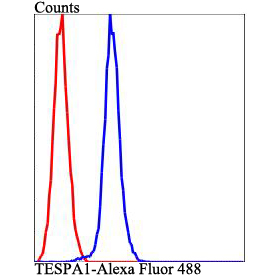
Fig7: Flow cytometric analysis of HepG2 cells with TESPA1 antibody at 1/100 dilution (blue) compared with an unlabelled control (cells without incubation with primary antibody; red). Alexa Fluor 488-conjugated goat anti-rabbit IgG was used as the secondar
- Background
Related Products / Services
Please note: All products are "FOR RESEARCH USE ONLY AND ARE NOT INTENDED FOR DIAGNOSTIC OR THERAPEUTIC USE"