


-
Product Name
Anti-SHH antibody
- Documents
-
Description
Rabbit monoclonal antibody to SHH
-
Tested applications
WB, ICC/IF, IHC-P, FC
-
Species reactivity
Human
-
Alternative names
TPT antibody; HHG1 antibody; HLP3 antibody; HPE3 antibody; SMMCI antibody; ShhNC antibody; TPTPS antibody; MCOPCB5 antibody
-
Isotype
Rabbit IgG
-
Preparation
This antigen of this antibody was recombinant protein
-
Clonality
Monoclonal
-
Formulation
Liquid, 1*TBS (pH7.4), 0.05% BSA, 40% Glycerol. Preservative: 0.05% Sodium Azide.
-
Storage instructions
Store at +4℃ after thawing. Aliquot store at -20℃ or -80℃. Avoid repeated freeze / thaw cycles.
-
Applications
WB: 1:1,000-1:2,000
ICC/IF: 1:50-1:200
IHC-P: 1:200-1:500
FC: 1:50-1:100
-
Validations
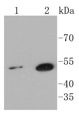
Fig1:; Western blot analysis of Sonic Hedgehog Protein on different lysates using anti-Sonic Hedgehog Protein antibody at 1/1,000 dilution.; Positive control:; Lane 1: Hela; Lane 2: HepG2
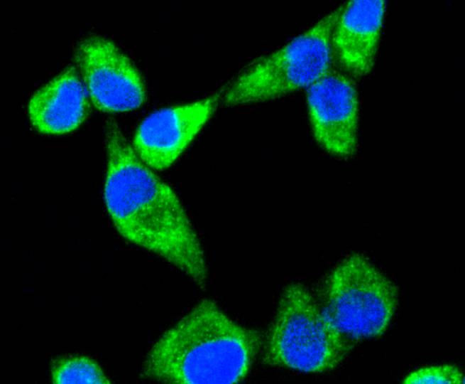
Fig2:; ICC staining Sonic Hedgehog Protein in Hela cells (green). The nuclear counter stain is DAPI (blue). Cells were fixed in paraformaldehyde, permeabilised with 0.25% Triton X100/PBS.
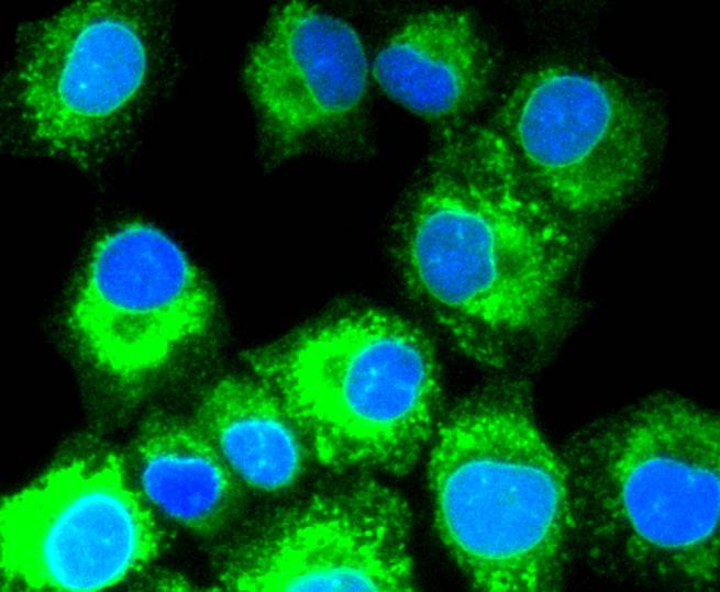
Fig3:; ICC staining Sonic Hedgehog Protein in A549 cells (green). The nuclear counter stain is DAPI (blue). Cells were fixed in paraformaldehyde, permeabilised with 0.25% Triton X100/PBS.
Fig4:; ICC staining Sonic Hedgehog Protein in 293 cells (green). The nuclear counter stain is DAPI (blue). Cells were fixed in paraformaldehyde, permeabilised with 0.25% Triton X100/PBS.
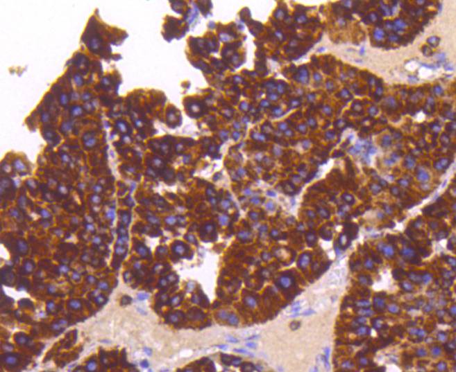
Fig5:; Immunohistochemical analysis of paraffin-embedded human liver cancer tissue using anti-Sonic Hedgehog Protein antibody. Counter stained with hematoxylin.
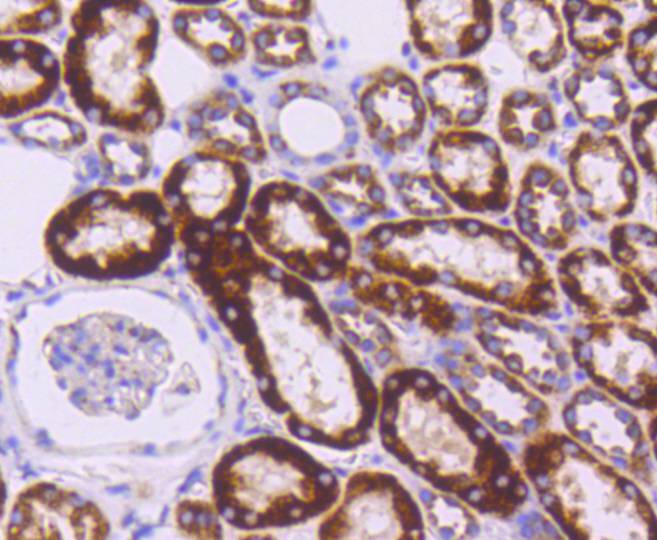
Fig6:; Immunohistochemical analysis of paraffin-embedded human kidney tissue using anti-Sonic Hedgehog Protein antibody. Counter stained with hematoxylin.
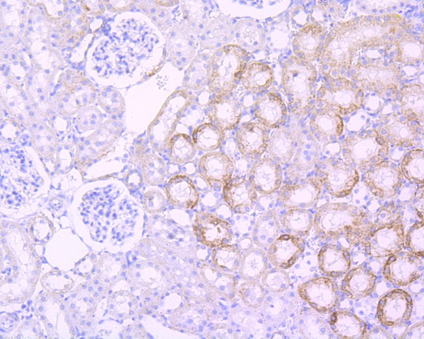
Fig7:; Immunohistochemical analysis of paraffin-embedded rat kidney tissue using anti-Sonic Hedgehog Protein antibody. Counter stained with hematoxylin.
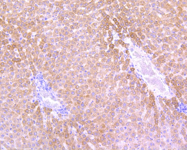
Fig8:; Immunohistochemical analysis of paraffin-embedded rat liver tissue using anti-Sonic Hedgehog Protein antibody. Counter stained with hematoxylin.
Fig9:; Flow cytometric analysis of Hela cells with Sonic Hedgehog Protein antibody at 1/50 dilution (red) compared with an unlabelled control (cells without incubation with primary antibody; black). Alexa Fluor 488-conjugated goat anti rabbit IgG was used as the secondary antibody.
- Background
-
References
- Zhang X et al. Expression of SOX9 and CDX2 in nongoblet columnar-lined esophagus predicts the detection of Barrett's esophagus during follow-up. Mod Pathol 28:654-61 (2015).
- Li H et al. Olfactomedin 4 deficiency promotes prostate neoplastic progression and is associated with upregulation of the hedgehog-signaling pathway. Sci Rep 5:16974 (2015).
Related Products / Services
Please note: All products are "FOR RESEARCH USE ONLY AND ARE NOT INTENDED FOR DIAGNOSTIC OR THERAPEUTIC USE"