
-
Product Name
Anti-NKX3-1 antibody
- Documents
-
Description
Mouse monoclonal antibody to NKX3-1
-
Tested applications
WB, IHC-P, FC
-
Species reactivity
Human
-
Alternative names
NKX3 antibody; BAPX2 antibody; NKX3A antibody; NKX3.1 antibody
-
Isotype
Mouse IgG2b
-
Preparation
This antigen of this antibody was recombinant protein
-
Clonality
Monoclonal
-
Formulation
Liquid, 1*TBS (pH7.4), 1%BSA, Preservative: 0.05% Sodium Azide.
-
Storage instructions
Store at +4℃ after thawing. Aliquot store at -20℃ or -80℃. Avoid repeated freeze / thaw cycles.
-
Applications
WB: 1:500-1:1,000
IHC-P: 1:50-1:200
FC: 1:100-1:200
-
Validations
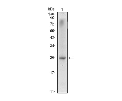
Fig1: Western blot analysis of NKX3A on LNCaP cell lysate using anti-NKX3A antibody at 1/1,000 dilution.
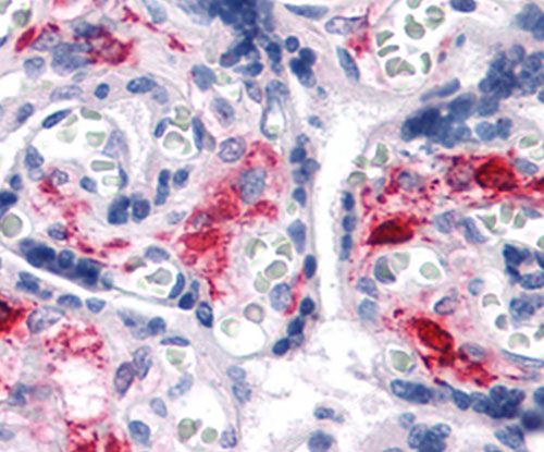
Fig2: Immunohistochemical analysis of paraffin-embedded human liver tissue using anti-NKX3A antibody. Counter stained with hematoxylin.
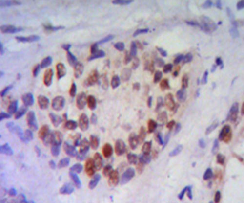
Fig3: Immunohistochemical analysis of paraffin-embedded human prostate tissue using anti-NKX3A antibody. Counter stained with hematoxylin.
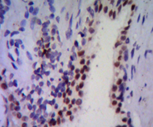
Fig4: Immunohistochemical analysis of paraffin-embedded human prostate tissue using anti-NKX3A antibody. Counter stained with hematoxylin.
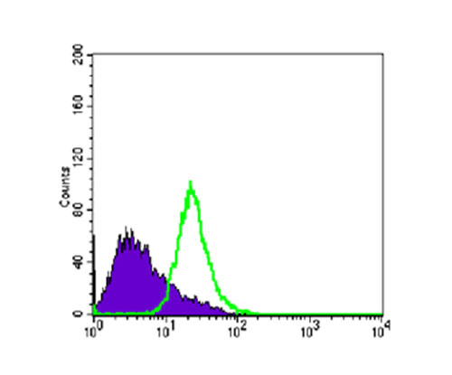
Fig5: Flow cytometric analysis of PC-3 cells with NKX3A antibody at 1/100 dilution (green) compared with an unlabelled control (cells without incubation with primary antibody; purple).
- Background
-
References
- Jiang A et al. p53 overexpression represses androgen-mediated induction of NKX3.1 in a prostate cancer cell line.Exp Mol Med 38(6):625-33 (2006).
- Zhang Y et al. Structural and functional analysis of domains mediating interaction between the bagpipe homologue, Nkx3.1 and serum response factor. Exp Biol Med (Maywood) 233(3):297-309 (2006).
Related Products / Services
Please note: All products are "FOR RESEARCH USE ONLY AND ARE NOT INTENDED FOR DIAGNOSTIC OR THERAPEUTIC USE"