
-
Product Name
Anti-LC3A/B Rabbit antibody
- Documents
-
Description
LC3A/B Rabbit polyclonal antibody
-
Tested applications
WB
-
Species reactivity
Human, Mouse, Rat
-
Alternative names
ATG8F; Autophagy related protein LC3 A; Autophagy related protein LC3 B; Autophagy related ubiquitin like modifier LC3 A; Autophagy related ubiquitin like modifier LC3 B; Autophagy-related protein LC3 B; Autophagy-related ubiquitin-like modifier LC3 B; LC antibody
-
Isotype
Rabbit IgG
-
Preparation
Antigen: Synthetic peptide.
-
Clonality
Polyclonal
-
Formulation
Purified Rabbit polyclonal antibody in PBS(pH 7.4) containing with 0.03% Proclin300 and 50% glycerol.
-
Storage instructions
Store at 4°C short term. Store at -20°C long term. Avoid freeze / thaw cycle.
-
Applications
WB: 1/1000
-
Validations
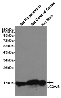
Western blot detection of LC3A/B in Rat Hippocampus, Rat Cerebral Cortex and Rat Brain lysates using LC3A/B Rabbit pAb (1:1000 diluted). Predicted band size: 15KDa. Observed band size:14, 16KDa.
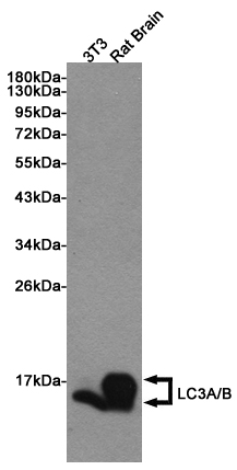
Western blot detection of LC3A/B in 3T3 and Rat Brain lysates using LC3A/B Rabbit pAb (1:1000 diluted). Predicted band size: 15KDa. Observed band size:14, 16KDa.
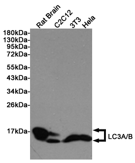
Western blot detection of LC3A/B in Rat Brain, C2C12, 3T3 and Hela cell lysates using LC3A/B Rabbit pAb (1:1000 diluted). Predicted band size: 15KDa. Observed band size:14, 16KDa.
-
Background
Swiss-Prot Acc.Q9H492,Q9GZQ8.Macroautophagy is the major inducible pathway for the general turnover of cytoplasmic constituents in eukaryotic cells, it is also responsible for the degradation of active cytoplasmic enzymes and organelles during nutrient starvation. Macroautophagy involves the formation of double-membrane bound autophagosomes which enclose the cytoplasmic constituent targeted for degradation in a membrane bound structure, which then fuse with the lysosome (or vacuole) releasing a single-membrane bound autophagic bodies which are then degraded within the lysosome (or vacuole). MAP1A and MAP1B are microtubule-associated proteins which mediate the physical interactions between microtubules and components of the cytoskeleton. These proteins are involved in formation of autophagosomal vacuoles (autophagosomes). MAP1A and MAP1B each consist of a heavy chain subunit and multiple light chain subunits. MAP1LC3a is one of the light chain subunits and can associate with either MAP1A or MAP1B. The precursor molecule is cleaved by APG4B/ATG4B to form the cytosolic form, LC3-I. This is activated by APG7L/ATG7, transferred to ATG3 and conjugated to phospholipid to form the membrane-bound form, LC3-II.
-
References
- Effect of metformin on human periodontal ligament stem cells cultured with polydopamine-templated hydroxyapatite
Related Products / Services
Please note: All products are "FOR RESEARCH USE ONLY AND ARE NOT INTENDED FOR DIAGNOSTIC OR THERAPEUTIC USE"