
-
Product Name
Anti-Eftud2 antibody
- Documents
-
Description
Mouse monoclonal antibody to Eftud2
-
Tested applications
WB, ICC, IHC-P, FC
-
Species reactivity
Human, Mouse, Rat
-
Alternative names
Snr antibody; 116kD antibody; 116kDa antibody; Snrp116 antibody; U5-116kD antibody
-
Isotype
Mouse IgG1
-
Preparation
This antigen of this antibody was peptide
-
Clonality
Monoclonal
-
Formulation
Liquid, 1*PBS (pH7.4), 0.2% BSA, 40% Glycerol. Preservative: 0.05% Sodium Azide.
-
Storage instructions
Store at +4℃ after thawing. Aliquot store at -20℃ or -80℃. Avoid repeated freeze / thaw cycles.
-
Applications
WB: 1:1,000-1:2,000
ICC: 1:50-1:200
IHC-P: 1:50-1:200
FC: 1:50-1:100
-
Validations
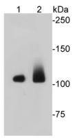
Fig1: Western blot analysis on different cell lysates using anti-EFTUD2 Mouse mAb. Positive control:; Lane 1: Jurkat; Lane 2: NIH/3T3
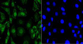
Fig2: Immunocytochemical staining of SH-SY-5Y cells using anti-EFTUD2 Mouse mAb
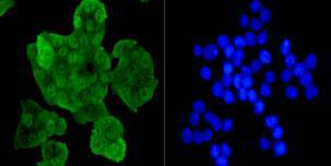
Fig3: Immunocytochemical staining of SW480 cells using anti-EFTUD2 Mouse mAb.
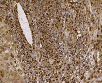
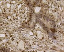
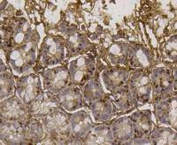
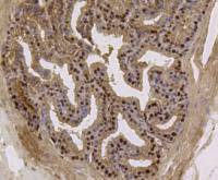
Fig4: Immunohistochemical analysis of paraffin- embedded human lung cancer tissue using anti-EFTUD2 Mouse mAb.
Fig5: Immunohistochemical analysis of paraffin- embedded human colon cancer tissue using anti-EFTUD2 Mouse mAb.
Fig6: Immunohistochemical analysis of paraffin- embedded mouse colon tissue using anti-EFTUD2 Mouse mAb.
Fig7: Immunohistochemical analysis of paraffin- embedded mouse prostate tissue using anti-EFTUD2 Mouse mAb.
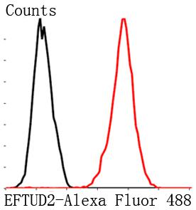
Fig8: Flow cytometric analysis of MCF-7 cells with EFTUD2 antibody at 1/100 dilution (red) compared with an unlabelled control (cells without ncubation with primary antibody; black). Goat anti mouse IgG (FITC) was used as the secondary antibody.
- Background
-
References
- Bian Y., Song C., Cheng K., et al. An enzyme assisted RP-RPLC approach for in-depth analysis of human liver phosphoproteome. J. Proteomics 96:253-262(2014).
- Horejsi Z., Stach L., Flower T.G., et al. Phosphorylation-dependent PIH1D1 interactions define substrate specificity of the R2TP cochaperone complex. Cell Rep. 7:19-26(2014).
Related Products / Services
Please note: All products are "FOR RESEARCH USE ONLY AND ARE NOT INTENDED FOR DIAGNOSTIC OR THERAPEUTIC USE"