
-
Product Name
Anti-Ccne1 antibody
- Documents
-
Description
Rabbit polyclonal antibody to Ccne1
-
Tested applications
WB, IHC-P, ICC/IF, FC
-
Species reactivity
Human, Mouse, Rat
-
Alternative names
Ccne antibody; CYCLE antibody
-
Isotype
Rabbit IgG
-
Preparation
This antigen of this antibody was klh conjugated synthetic peptide derived from rat cyclin e 375-411/411
-
Clonality
Polyclonal
-
Formulation
Liquid, 0.01M TBS(pH7.4) with 1% BSA, 0.03% Proclin300 and 50% Glycerol.
-
Storage instructions
Store at -20℃ for one year. Avoid repeated freeze/thaw cycles. The lyophilized antibody is stable at room temperature for at least one month and for greater than a year when kept at -20℃. When reconstituted in sterile pH 7.4 0.01M PBS or diluent of antibody the antibody is stable for at least two weeks at 2-4℃.
-
Applications
WB:1:500-2000
IHC-P:1:400-800
FC:1μg/Test
IF:1:100-500
-
Validations
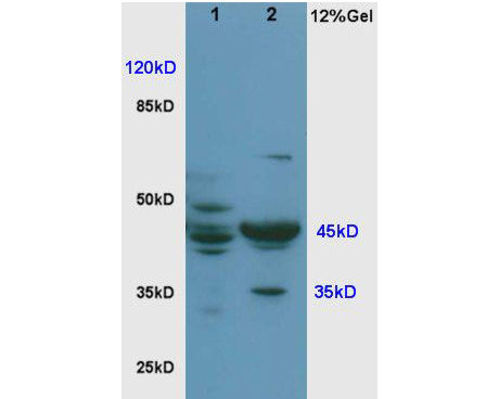
Fig1: Sample:; Brain(Rat) lysate at30ug;; Lung(Rat) lysate at 30ug;; Primary: Anti-Cyclin E at 1:200;; Secondary: HRP conjugated Goat-Anti-Rabbit IgG(bse-0295G) at 1: 3000;; Predicted band size : 45kD; Observed band size : 45kD
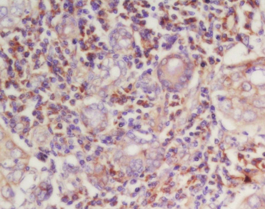
Fig2: Tissue/cell: human laryngocarcinoma; 4% Paraformaldehyde-fixed and paraffin-embedded;; Antigen retrieval: citrate buffer ( 0.01M, pH 6.0 ), Boiling bathing for 15min; Block endogenous peroxidase by 3% Hydrogen peroxide for 30min; Blocking buffer (normal goat serum,C-0005) at 37℃ for 20 min;; Incubation: Anti-Cyclin-E Polyclonal Antibody, Unconjugated 1:200, overnight at 4℃, followed by conjugation to the secondary antibody(SP-0023) and DAB(C-0010) staining
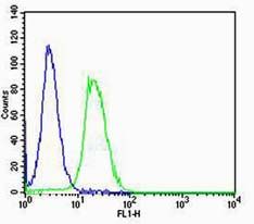
Fig3: Cell: NIH/3T3; Concentration:1:100; Host/Isotype:Rabbit/IgG; Flow cytometric analysis of primary antibody (Cat#: 175357#) on NIH/3T3(green) compared with Rabbit IgG isotype control in the absence of primary antibody (blue) followed by Alexa Fluor 488-conjugated goat anti-rabbit IgG(H+L) secondary antibody .
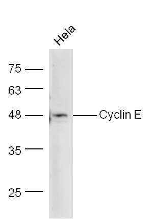
Fig4: Sample: Hela Lysate at 40 ug; Primary: Anti-Cyclin E at 1/300 dilution; Secondary: IRDye800CW Goat Anti-Rabbit IgG at 1/10000 dilution; Predicted band size: 45 kD; Observed band size: 48 kD
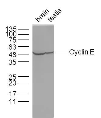
Fig5: Sample:; Brain(Mouse) Lysate at 40 ug; Testis(Mouse) Lysate at 40 ug; Primary: Anti-Cyclin E at 1/300 dilution; Secondary: IRDye800CW Goat Anti-Rabbit IgG at 1/10000 dilution; Predicted band size: 45 kD; Observed band size: 48 kD
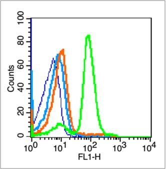
Fig6: Blank control (blue line): Mouse spleen cells (blue).; Primary Antibody (green line): Rabbit Anti-Cyclin E1 antibody ; Dilution: 1μg /10^6 cells;; Isotype Control Antibody (orange line): Rabbit IgG .; Secondary Antibody (white blue line): Goat anti-rabbit IgG-FITC; Dilution: 1μg /test.; Protocol; The cells were fixed with 70% ethanol (overninght at 4℃) and then permeabilized with 0.1% PBS-Tween for 20 min at room temperature. Cells stained with Primary Antibody for 30 min at room temperature. The cells were then incubated in 1 X PBS/2%BSA/10% goat serum to block non-specific protein-protein interactions followed by the antibody for 15 min at room temperature. The secondary antibody used for 40 min at room temperature. Acquisition of 20,000 events was performed.
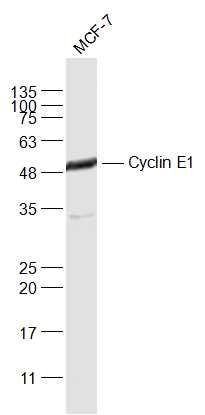
Fig7: Sample:; MCF-7(Human) Cell Lysate at 30 ug; Primary: Anti-Cyclin E1 at 1/1000 dilution; Secondary: IRDye800CW Goat Anti-Rabbit IgG at 1/20000 dilution; Predicted band size: 45 kD; Observed band size: 48 kD
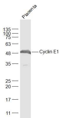
Fig8: Sample:; Placenta (Mouse) Lysate at 40 ug; Primary: Anti-Cyclin E1 at 1/1000 dilution; Secondary: IRDye800CW Goat Anti-Rabbit IgG at 1/20000 dilution; Predicted band size: 45 kD; Observed band size: 48 kD
- Background
Related Products / Services
Please note: All products are "FOR RESEARCH USE ONLY AND ARE NOT INTENDED FOR DIAGNOSTIC OR THERAPEUTIC USE"