
-
Product Name
WDR26 Polyclonal Antibody
- Documents
-
Description
Polyclonal antibody to WDR26
-
Tested applications
WB
-
Species reactivity
Human, Mouse, Rat
-
Alternative names
WDR26 antibody; CDW2 antibody; GID7 antibody; MIP2 antibody; WD repeat domain 26 antibody
-
Isotype
Rabbit IgG
-
Preparation
Antigen: Recombinant fusion protein containing a sequence corresponding to amino acids 362-661 of human WDR26 (NP_079436.4).
-
Clonality
Polyclonal
-
Formulation
PBS with 0.02% sodium azide, 50% glycerol, pH7.3.
-
Storage instructions
Store at -20℃. Avoid freeze / thaw cycles.
-
Applications
WB 1:1000 - 1:2000
-
Validations
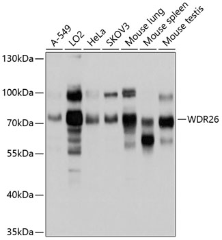
Western blot - WDR26 Polyclonal Antibody
Western blot analysis of extracts of various cell lines, using WDR26 antibody at 1:1000 dilution.Secondary antibody: HRP Goat Anti-Rabbit IgG (H+L) at 1:10000 dilution.Lysates/proteins: 25ug per lane.Blocking buffer: 3% nonfat dry milk in TBST.Detection: ECL Basic Kit .Exposure time: 5s.
-
Background
G-beta-like protein involved in cell signal transduction. Acts as a negative regulator in MAPK signaling pathway. Functions as a scaffolding protein to promote G beta:gamma-mediated PLCB2 plasma membrane translocation and subsequent activation in leukocytes. Core component of the CTLH E3 ubiquitin-protein ligase complex that selectively accepts ubiquitin from UBE2H and mediates ubiquitination and subsequent proteasomal degradation of the transcription factor HBP1. Acts as a negative regulator of the canonical Wnt signaling pathway through preventing ubiquitination of beta-catenin CTNNB1 by the beta-catenin destruction complex, thus negatively regulating CTNNB1 degradation. Serves as a scaffold to coordinate PI3K/AKT pathway-driven cell growth and migration. Protects cells from oxidative stress-induced apoptosis via the down-regulation of AP-1 transcriptional activity as well as by inhibiting cytochrome c release from mitochondria. Protects also cells by promoting hypoxia-mediated autophagy and mitophagy (By similarity).
Related Products / Services
Please note: All products are "FOR RESEARCH USE ONLY AND ARE NOT INTENDED FOR DIAGNOSTIC OR THERAPEUTIC USE"