
-
Product Name
TDP-43 (human specific) antibody
- Documents
-
Description
TDP-43 (human specific) Mouse Monoclonal antibody. Positive IHC detected in human brain(FTLD-U) tissue, human brain tissue, human gliomas tissue, human pancreas cancer tissue, human pancreas tissue. Positive FC detected in MCF-7 cells. Positive WB detected in K-562 cells, human brain tissue, human heart tissue, human placenta tissue. Positive IP detected in K-562 cells. Observed molecular weight by Western-blot: 43 kDa
-
Tested applications
ELISA, IP, FC, WB, IHC
-
Species reactivity
Human; other species not tested.
-
Alternative names
ALS10 antibody; TAR DNA binding protein antibody; TAR DNA binding protein 43 antibody; TARDBP antibody; TDP 43 antibody; TDP43 antibody
-
Isotype
Mouse IgG1
-
Preparation
This antibody was obtained by immunization of TDP-43 (human specific) recombinant protein (Accession Number: BC001487). Purification method: Caprylic acid/ammonium sulfate precipitation.
-
Clonality
Monoclonal
-
Formulation
PBS with 0.1% sodium azide and 50% glycerol pH 7.3.
-
Storage instructions
Store at -20℃. DO NOT ALIQUOT
-
Applications
Recommended Dilution:
WB: 1:5000-1:50000
IP: 1:500-1:5000
IHC: N/A
-
Validations
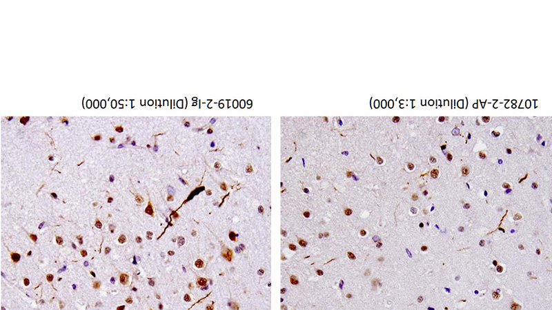
40X of FTLD-U case stained by Catalog No:115925 and Catalog No:107618, showing dystrophic neurites. (Figs were provided by Linda K. Kwong)
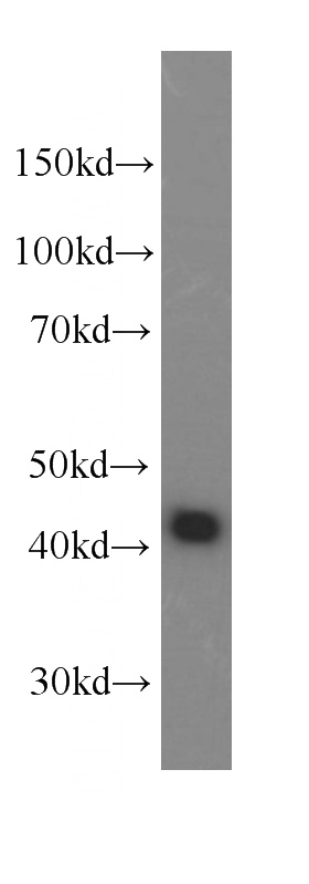
K-562 cells were subjected to SDS PAGE followed by western blot with Catalog No:107618(TDP-43 Antibody) at dilution of 1:10000
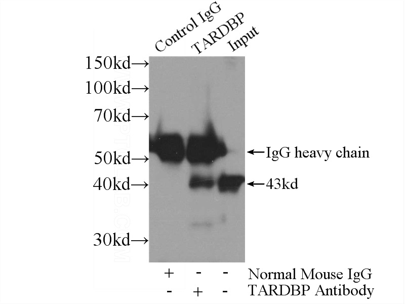
IP Result of anti-TDP-43 (IP:Catalog No:107618, 5ug; Detection:Catalog No:107618 1:1000) with K-562 cells lysate 1720ug.
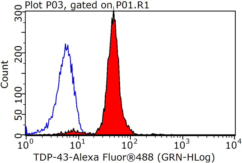
1X10^6 MCF-7 cells were stained with 0.2ug TDP-43 antibody (Catalog No:107618, red) and control antibody (blue). Fixed with 90% MeOH blocked with 3% BSA (30 min). Alexa Fluor 488-congugated AffiniPure Goat Anti-Mouse IgG(H+L) with dilution 1:1500.
-
Background
Transactivation response (TAR) DNA-binding protein of 43 kDa (also known as TARDBP or TDP-43) was first isolated as a transcriptional inactivator binding to the TAR DNA element of the HIV-1 virus. Neumann et al. (2006) found that a hyperphosphorylated, ubiquitinated, and cleaved form of TARDBP, known as pathologic TDP-43, is the major component of the tau-negative and ubiquitin-positive inclusions that characterize amyotrophic lateral sclerosis (ALS) and the most common pathological subtype of frontotemporal lobar degeneration (FTLD-U). 60019-2-Ig is a mouse monoclonal antibody recognizing the cleavage product of 20-30 kDa in addition to the native and phosphorylated forms of TDP-43. Immunohistochemical analyses of TDP-43 using this antibody detect both normal diffuse nuclear staining and insoluble inclusions in pathologic tissues. The epitope of 60019-2-Ig has been determined to locate at residues 203-209 (TEDELRE) by Hiroshi Tsuji et al. (2012), which is involved in the formation of pathologic TDP-43 aggregation. Notably this antibody only recognizes human TDP-43 but not reacts with mouse or rat TDP-43.
-
References
- Lee EB, Lee VM, Trojanowski JQ, Neumann M. TDP-43 immunoreactivity in anoxic, ischemic and neoplastic lesions of the central nervous system. Acta neuropathologica. 115(3):305-11. 2008.
- Rohn TT. Caspase-cleaved TAR DNA-binding protein-43 is a major pathological finding in Alzheimer's disease. Brain research. 1228:189-98. 2008.
- Deng HX, Zhai H, Bigio EH. FUS-immunoreactive inclusions are a common feature in sporadic and non-SOD1 familial amyotrophic lateral sclerosis. Annals of neurology. 67(6):739-48. 2010.
- Deng HX, Chen W, Hong ST. Mutations in UBQLN2 cause dominant X-linked juvenile and adult-onset ALS and ALS/dementia. Nature. 477(7363):211-5. 2011.
- Necchi D, Lomoio S, Scherini E. Dysfunction of the ubiquitin-proteasome system in the cerebellum of aging Ts65Dn mice. Experimental neurology. 232(2):114-8. 2011.
- Tsuji H, Nonaka T, Yamashita M. Epitope mapping of antibodies against TDP-43 and detection of protease-resistant fragments of pathological TDP-43 in amyotrophic lateral sclerosis and frontotemporal lobar degeneration. Biochemical and biophysical research communications. 417(1):116-21. 2012.
- Cohen TJ, Hwang AW, Unger T, Trojanowski JQ, Lee VM. Redox signalling directly regulates TDP-43 via cysteine oxidation and disulphide cross-linking. The EMBO journal. 31(5):1241-52. 2012.
- Inamori Y, Higuchi I, Inoue T. Inclusion body myositis coexisting with hypertrophic cardiomyopathy: an autopsy study. Neuromuscular disorders : NMD. 22(8):747-54. 2012.
Related Products / Services
Please note: All products are "FOR RESEARCH USE ONLY AND ARE NOT INTENDED FOR DIAGNOSTIC OR THERAPEUTIC USE"