
-
Product Name
RIPK1 Polyclonal Antibody
- Documents
-
Description
Polyclonal antibody to RIPK1
-
Tested applications
WB, IHC, IF, IP
-
Species reactivity
Human, Mouse, Rat
-
Alternative names
RIPK1 antibody; RIP antibody; RIP-1 antibody; RIP1 antibody; receptor interacting serine/threonine kinase 1 antibody
-
Isotype
Rabbit IgG
-
Preparation
Antigen: Recombinant fusion protein containing a sequence corresponding to amino acids 170-440 of human RIPK1 (NP_003795.2).
-
Clonality
Polyclonal
-
Formulation
PBS with 0.02% sodium azide, 50% glycerol, pH7.3.
-
Storage instructions
Store at -20℃. Avoid freeze / thaw cycles.
-
Applications
WB 1:500 - 1:2000
IHC 1:50 - 1:200
IF 1:50 - 1:200
IP 1:50 - 1:200 -
Validations
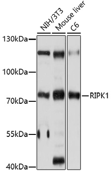
Western blot - RIPK1 Polyclonal Antibody
Western blot analysis of extracts of various cell lines,using RIPK1 antibody at 1:1000 dilution.Secondary antibody: HRP Goat Anti-Rabbit IgG (H+L) at 1:10000 dilution.Lysates/proteins: 25ug per lane.Blocking buffer: 3% nonfat dry milk in TBST.Detection: ECL Basic Kit .Exposure time: 10s.
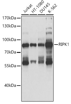
Western blot - RIPK1 Polyclonal Antibody
Western blot analysis of extracts of various cell lines, using RIPK1 antibody at 1:1000 dilution.Secondary antibody: HRP Goat Anti-Rabbit IgG (H+L) at 1:10000 dilution.Lysates/proteins: 25ug per lane.Blocking buffer: 3% nonfat dry milk in TBST.Detection: ECL Basic Kit .Exposure time: 1s.
Immunohistochemistry - RIPK1 Polyclonal Antibody
Immunohistochemistry of paraffin-embedded human tonsil using RIPK1 antibody at dilution of 1:100 (40x lens).
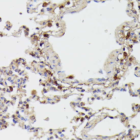
Immunohistochemistry - RIPK1 Polyclonal Antibody
Immunohistochemistry of paraffin-embedded rat lung using RIPK1 antibody at dilution of 1:100 (40x lens).
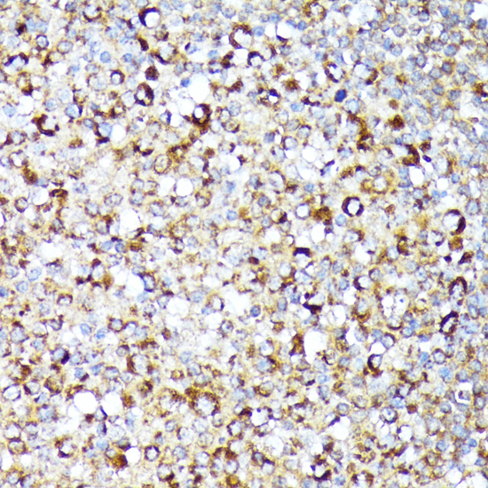
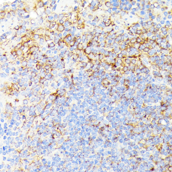
Immunohistochemistry - RIPK1 Polyclonal Antibody
Immunohistochemistry of paraffin-embedded mouse spleen using RIPK1 antibody at dilution of 1:100 (40x lens).
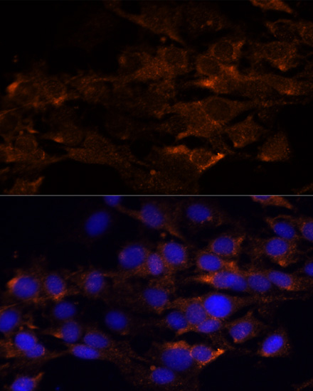
Immunofluorescence - RIPK1 Polyclonal Antibody
Immunofluorescence analysis of C6 cells using RIPK1 antibody at dilution of 1:100. Blue: DAPI for nuclear staining.
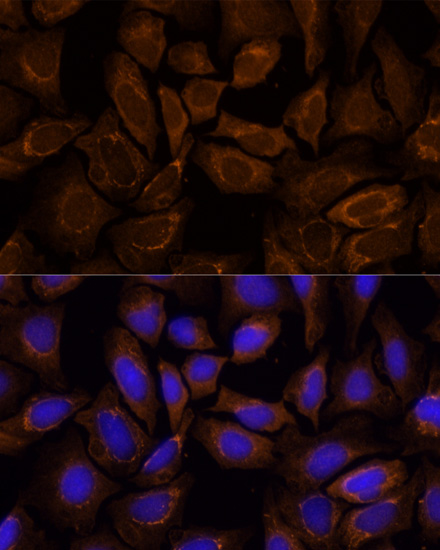
Immunofluorescence - RIPK1 Polyclonal Antibody
Immunofluorescence analysis of U-2 OS cells using RIPK1 antibody at dilution of 1:100. Blue: DAPI for nuclear staining.
-
Background
Serine-threonine kinase which transduces inflammatory and cell-death signals (programmed necrosis) following death receptors ligation, activation of pathogen recognition receptors (PRRs), and DNA damage. Upon activation of TNFR1 by the TNF-alpha family cytokines, TRADD and TRAF2 are recruited to the receptor. Phosphorylates DAB2IP at 'Ser-728' in a TNF-alpha-dependent manner, and thereby activates the MAP3K5-JNK apoptotic cascade. Ubiquitination by TRAF2 via 'Lys-63'-link chains acts as a critical enhancer of communication with downstream signal transducers in the mitogen-activated protein kinase pathway and the NF-kappa-B pathway, which in turn mediate downstream events including the activation of genes encoding inflammatory molecules. Polyubiquitinated protein binds to IKBKG/NEMO, the regulatory subunit of the IKK complex, a critical event for NF-kappa-B activation. Interaction with other cellular RHIM-containing adapters initiates gene activation and cell death. RIPK1 and RIPK3 association, in particular, forms a necrosis-inducing complex.
Related Products / Services
Please note: All products are "FOR RESEARCH USE ONLY AND ARE NOT INTENDED FOR DIAGNOSTIC OR THERAPEUTIC USE"