-
Product Name
PMAIP1 Polyclonal Antibody
- Documents
-
Description
Polyclonal antibody to PMAIP1
-
Tested applications
WB, IF
-
Species reactivity
Human, Mouse, Rat
-
Alternative names
PMAIP1 antibody; APR antibody; NOXA antibody; phorbol-12-myristate-13-acetate-induced protein 1 antibody
-
Isotype
Rabbit IgG
-
Preparation
Antigen: A synthetic peptide corresponding to a sequence within amino acids 1-54 of human PMAIP1 (NP_066950.1).
-
Clonality
Polyclonal
-
Formulation
PBS with 0.02% sodium azide, 50% glycerol, pH7.3.
-
Storage instructions
Store at -20℃. Avoid freeze / thaw cycles.
-
Applications
WB 1:500 - 1:2000
IF 1:50 - 1:200 -
Validations
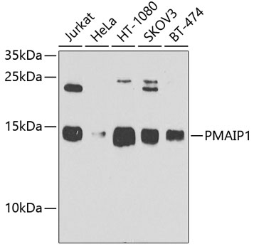
Western blot - PMAIP1 Polyclonal Antibody
Western blot analysis of extracts of various cell lines, using PMAIP1 antibody at 1:1000 dilution.Secondary antibody: HRP Goat Anti-Rabbit IgG (H+L) at 1:10000 dilution.Lysates/proteins: 25ug per lane.Blocking buffer: 3% nonfat dry milk in TBST.Detection: ECL Basic Kit .Exposure time: 90s.
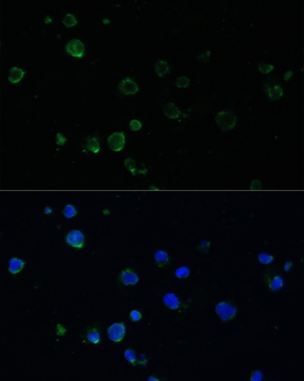
Immunofluorescence - PMAIP1 Polyclonal Antibody
Immunofluorescence analysis of Jurkat cells using PMAIP1 antibody at dilution of 1:100. Blue: DAPI for nuclear staining.
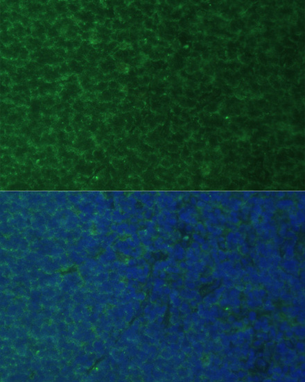
Immunofluorescence - PMAIP1 Polyclonal Antibody
Immunofluorescence analysis of rat breast cells using PMAIP1 antibody at dilution of 1:100. Blue: DAPI for nuclear staining.
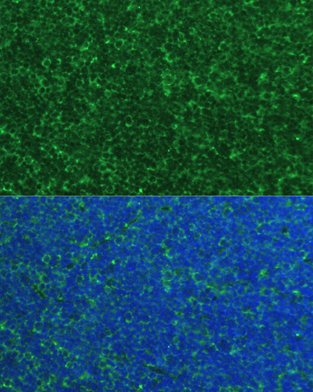
Immunofluorescence - PMAIP1 Polyclonal Antibody
Immunofluorescence analysis of mouse breast cells using PMAIP1 antibody at dilution of 1:100. Blue: DAPI for nuclear staining.
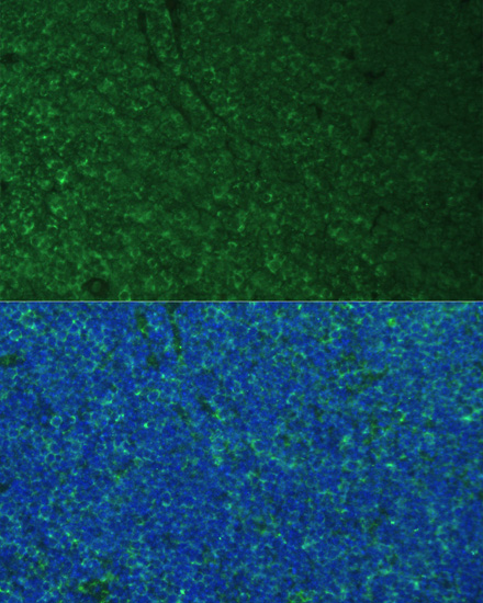
Immunofluorescence - PMAIP1 Polyclonal Antibody
Immunofluorescence analysis of mouse breast cells using PMAIP1 antibody at dilution of 1:100. Blue: DAPI for nuclear staining.
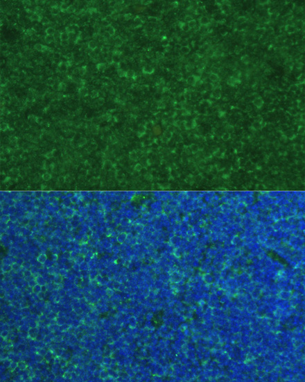
Immunofluorescence - PMAIP1 Polyclonal Antibody
Immunofluorescence analysis of mouse breast cells using PMAIP1 antibody at dilution of 1:100. Blue: DAPI for nuclear staining.
-
Background
Promotes activation of caspases and apoptosis. Promotes mitochondrial membrane changes and efflux of apoptogenic proteins from the mitochondria. Contributes to p53/TP53-dependent apoptosis after radiation exposure. Promotes proteasomal degradation of MCL1. Competes with BAK1 for binding to MCL1 and can displace BAK1 from its binding site on MCL1 (By similarity). Competes with BIM/BCL2L11 for binding to MCL1 and can displace BIM/BCL2L11 from its binding site on MCL1.
Related Products / Services
Please note: All products are "FOR RESEARCH USE ONLY AND ARE NOT INTENDED FOR DIAGNOSTIC OR THERAPEUTIC USE"
