
-
Product Name
PKD2 Polyclonal Antibody
- Documents
-
Description
Polyclonal antibody to PKD2
-
Tested applications
WB
-
Species reactivity
Human, Mouse, Rat
-
Alternative names
PKD2 antibody; APKD2 antibody; PC2 antibody; PKD4 antibody; Pc-2 antibody; TRPP2 antibody; polycystin-2 antibody
-
Isotype
Rabbit IgG
-
Preparation
Antigen: Recombinant fusion protein containing a sequence corresponding to amino acids 240-410 of human PKD2 (NP_000288.1).
-
Clonality
Polyclonal
-
Formulation
PBS with 0.02% sodium azide, 50% glycerol, pH7.3.
-
Storage instructions
Store at -20℃. Avoid freeze / thaw cycles.
-
Applications
WB 1:500 - 1:1000
-
Validations
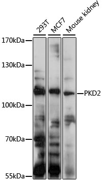
Western blot - PKD2 Polyclonal Antibody
Western blot analysis of extracts of various cell lines, using PKD2 antibody at 1:1000 dilution.Secondary antibody: HRP Goat Anti-Rabbit IgG (H+L) at 1:10000 dilution.Lysates/proteins: 25ug per lane.Blocking buffer: 3% nonfat dry milk in TBST.Detection: ECL Basic Kit .Exposure time: 30s.
-
Background
Component of a heteromeric calcium-permeable ion channel formed by PKD1 and PKD2 that is activated by interaction between PKD1 and a Wnt family member, such as WNT3A and WNT9B. Can also form a functional, homotetrameric ion channel. Functions as a cation channel involved in fluid-flow mechanosensation by the primary cilium in renal epithelium. Functions as outward-rectifying K(+) channel, but is also permeable to Ca(2+), and to a much lesser degree also to Na(+). May contribute to the release of Ca(2+) stores from the endoplasmic reticulum. Together with TRPV4, forms mechano- and thermosensitive channels in cilium. PKD1 and PKD2 may function through a common signaling pathway that is necessary to maintain the normal, differentiated state of renal tubule cells. Acts as a regulator of cilium length, together with PKD1. The dynamic control of cilium length is essential in the regulation of mechanotransductive signaling. The cilium length response creates a negative feedback loop whereby fluid shear-mediated deflection of the primary cilium, which decreases intracellular cAMP, leads to cilium shortening and thus decreases flow-induced signaling. Also involved in left-right axis specification via its role in sensing nodal flow; forms a complex with PKD1L1 in cilia to facilitate flow detection in left-right patterning. Detection of asymmetric nodal flow gives rise to a Ca(2+) signal that is required for normal, asymmetric expression of genes involved in the specification of body left-right laterality (By similarity).
Related Products / Services
Please note: All products are "FOR RESEARCH USE ONLY AND ARE NOT INTENDED FOR DIAGNOSTIC OR THERAPEUTIC USE"