

-
Product Name
PIK3C2A Polyclonal Antibody
- Documents
-
Description
Polyclonal antibody to PIK3C2A
-
Tested applications
WB, IHC, IF, IP
-
Species reactivity
Human, Mouse, Rat
-
Alternative names
PIK3C2A antibody; CPK antibody; PI3-K-C2 antibody; PI3-K-C2A antibody; PI3K-C2-alpha antibody; PI3K-C2alpha antibody; phosphatidylinositol-4-phosphate 3-kinase catalytic subunit type 2 alpha antibody
-
Isotype
Rabbit IgG
-
Preparation
Antigen: Recombinant fusion protein containing a sequence corresponding to amino acids 1-200 of human PIK3C2A (NP_002636.2).
-
Clonality
Polyclonal
-
Formulation
PBS with 0.02% sodium azide, 50% glycerol, pH7.3.
-
Storage instructions
Store at -20℃. Avoid freeze / thaw cycles.
-
Applications
WB 1:500 - 1:2000
IHC 1:50 - 1:100
IF 1:50 - 1:100
IP 1:50 - 1:100 -
Validations
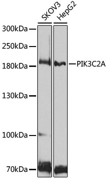
Western blot - PIK3C2A Polyclonal Antibody
Western blot analysis of extracts of various cell lines, using PIK3C2A antibody at 1:1000 dilution.Secondary antibody: HRP Goat Anti-Rabbit IgG (H+L) at 1:10000 dilution.Lysates/proteins: 25ug per lane.Blocking buffer: 3% nonfat dry milk in TBST.Detection: ECL Basic Kit .Exposure time: 90s.
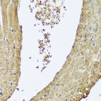
Immunohistochemistry - PIK3C2A Polyclonal Antibody
Immunohistochemistry of paraffin-embedded mouse heart using PIK3C2A antibody at dilution of 1:100 (40x lens).
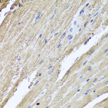
Immunohistochemistry - PIK3C2A Polyclonal Antibody
Immunohistochemistry of paraffin-embedded rat heart using PIK3C2A antibody at dilution of 1:100 (40x lens).
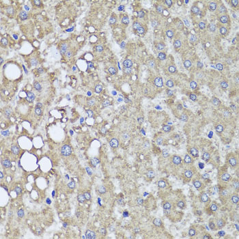
Immunohistochemistry - PIK3C2A Polyclonal Antibody
Immunohistochemistry of paraffin-embedded human liver injury using PIK3C2A antibody at dilution of 1:100 (40x lens).
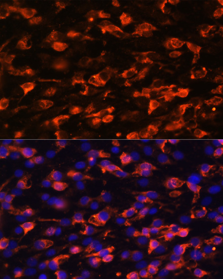
Immunofluorescence - PIK3C2A Polyclonal Antibody
Immunofluorescence analysis of C6 cells using PIK3C2A antibody at dilution of 1:100. Blue: DAPI for nuclear staining.
Immunoprecipitation - PIK3C2A Polyclonal Antibody
Immunoprecipitation analysis of 200ug extracts of HeLa cells, using 3 ug PIK3C2A antibody . Western blot was performed from the immunoprecipitate using PIK3C2A antibody at a dilition of 1:1000.
-
Background
Generates phosphatidylinositol 3-phosphate (PtdIns3P) and phosphatidylinositol 3,4-bisphosphate (PtdIns(3,4)P2) that act as second messengers. Has a role in several intracellular trafficking events. Functions in insulin signaling and secretion. Required for translocation of the glucose transporter SLC2A4/GLUT4 to the plasma membrane and glucose uptake in response to insulin-mediated RHOQ activation. Regulates insulin secretion through two different mechanisms: involved in glucose-induced insulin secretion downstream of insulin receptor in a pathway that involves AKT1 activation and TBC1D4/AS160 phosphorylation, and participates in the late step of insulin granule exocytosis probably in insulin granule fusion. Synthesizes PtdIns3P in response to insulin signaling. Functions in clathrin-coated endocytic vesicle formation and distribution. Regulates dynamin-independent endocytosis, probably by recruiting EEA1 to internalizing vesicles. In neurosecretory cells synthesizes PtdIns3P on large dense core vesicles. Participates in calcium induced contraction of vascular smooth muscle by regulating myosin light chain (MLC) phosphorylation through a mechanism involving Rho kinase-dependent phosphorylation of the MLCP-regulatory subunit MYPT1. May play a role in the EGF signaling cascade. May be involved in mitosis and UV-induced damage response. Required for maintenance of normal renal structure and function by supporting normal podocyte function.
Related Products / Services
Please note: All products are "FOR RESEARCH USE ONLY AND ARE NOT INTENDED FOR DIAGNOSTIC OR THERAPEUTIC USE"