-
Product Name
PDE4DIP Polyclonal Antibody
- Documents
-
Description
Polyclonal antibody to PDE4DIP
-
Tested applications
WB, IHC
-
Species reactivity
Human, Mouse, Rat
-
Alternative names
PDE4DIP antibody; CMYA2 antibody; MMGL antibody; myomegalin antibody
-
Isotype
Rabbit IgG
-
Preparation
Antigen: Recombinant fusion protein containing a sequence corresponding to amino acids 1-310 of human PDE4DIP (NP_071754.3).
-
Clonality
Polyclonal
-
Formulation
PBS with 0.02% sodium azide, 50% glycerol, pH7.3.
-
Storage instructions
Store at -20℃. Avoid freeze / thaw cycles.
-
Applications
WB 1:500 - 1:2000
IHC 1:50 - 1:200 -
Validations

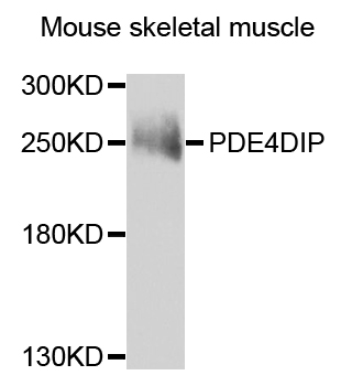
Western blot - PDE4DIP Polyclonal Antibody
Western blot analysis of extracts of mouse skeletal muscle, using PDE4DIP antibody at 1:1000 dilution.Secondary antibody: HRP Goat Anti-Rabbit IgG (H+L) at 1:10000 dilution.Lysates/proteins: 25ug per lane.Blocking buffer: 3% nonfat dry milk in TBST.Detection: ECL Enhanced Kit .Exposure time: 60s.

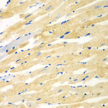
Immunohistochemistry - PDE4DIP Polyclonal Antibody
Immunohistochemistry of paraffin-embedded rat heart using PDE4DIP antibody at dilution of 1:100 (40x lens).

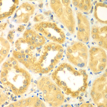
Immunohistochemistry - PDE4DIP Polyclonal Antibody
Immunohistochemistry of paraffin-embedded human kidney using PDE4DIP antibody at dilution of 1:100 (40x lens).

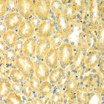
Immunohistochemistry - PDE4DIP Polyclonal Antibody
Immunohistochemistry of paraffin-embedded mouse kidney using PDE4DIP antibody at dilution of 1:100 (40x lens).
-
Background
Functions as an anchor sequestering components of the cAMP-dependent pathway to Golgi and/or centrosomes (By similarity).; Isoform 13: Participates in microtubule dynamics, promoting microtubule assembly. Depending upon the cell context, may act at the level of the Golgi apparatus or that of the centrosome. In complex with AKAP9, recruits CAMSAP2 to the Golgi apparatus and tethers non-centrosomal minus-end microtubules to the Golgi, an important step for polarized cell movement. In complex with AKAP9, EB1/MAPRE1 and CDK5RAP2, contributes to microtubules nucleation and extension from the centrosome to the cell periphery, a crucial process for directed cell migration, mitotic spindle orientation and cell-cycle progression.
Related Products / Services
Please note: All products are "FOR RESEARCH USE ONLY AND ARE NOT INTENDED FOR DIAGNOSTIC OR THERAPEUTIC USE"
