
-
Product Name
nucleobindin 1 antibody
- Documents
-
Description
nucleobindin 1 Rabbit Polyclonal antibody. Positive IF detected in HepG2 cells. Positive FC detected in HepG2 cells. Positive IP detected in HepG2 cells. Positive WB detected in MCF7 cells, A431 cells, HeLa cells, HepG2 cells. Observed molecular weight by Western-blot: 63 kDa
-
Tested applications
ELISA, WB, IF, FC, IP
-
Species reactivity
Human,Mouse,Rat; other species not tested.
-
Alternative names
CALNUC antibody; DKFZp686A15286 antibody; FLJ40471 antibody; NUC antibody; NUCB1 antibody; nucleobindin 1 antibody
-
Isotype
Rabbit IgG
-
Preparation
This antibody was obtained by immunization of nucleobindin 1 recombinant protein (Accession Number: NM_006184). Purification method: Antigen affinity purified.
-
Clonality
Polyclonal
-
Formulation
PBS with 0.1% sodium azide and 50% glycerol pH 7.3.
-
Storage instructions
Store at -20℃. DO NOT ALIQUOT
-
Applications
Recommended Dilution:
WB: 1:200-1:2000
IP: 1:500-1:5000
IF: 1:20-1:200
-
Validations
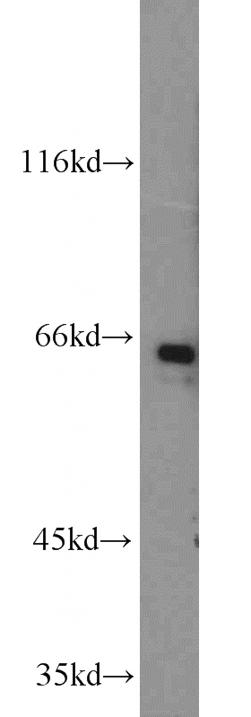
MCF7 cells were subjected to SDS PAGE followed by western blot with Catalog No:113408(NUCB1 antibody) at dilution of 1:1000
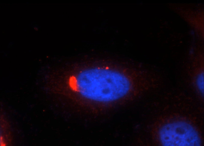
Immunofluorescent analysis of HepG2 cells, using NUCB1 antibody Catalog No:113408 at 1:50 dilution and Rhodamine-labeled goat anti-rabbit IgG (red). Blue pseudocolor = DAPI (fluorescent DNA dye).
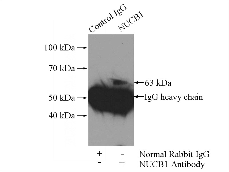
IP Result of anti-NUCB1 (IP:Catalog No:113408, 4ug; Detection:Catalog No:113408 1:1000) with HepG2 cells lysate 2400ug.
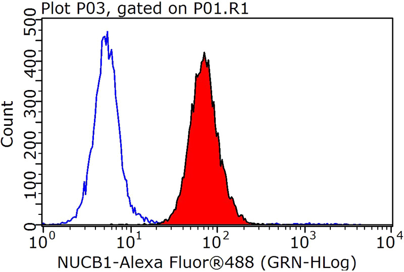
1X10^6 HepG2 cells were stained with 0.2ug NUCB1 antibody (Catalog No:113408, red) and control antibody (blue). Fixed with 90% MeOH blocked with 3% BSA (30 min). Alexa Fluor 488-congugated AffiniPure Goat Anti-Rabbit IgG(H+L) with dilution 1:1000.
-
Background
Nucleobindin (Nuc) was first identified as a secreted protein of 55 kDa that promotes production of DNA-specific antibodies in lupus-prone MRL/lpr mice. Nuc contains a signal peptide, a DNA-binding site, two calcium-binding motifs (EF-hand motifs), and a leucine zipper. Nucleobindin is found in both cytosol and membrane and is localized to cis-Golgi cisternae and the cis-Golgi network (CGN). Nucleobindin is involved in autoimmunity, apoptosis and calcium homeostasis in the bone matrix. NUC was located at human chromosome 19q13.2-q13.4.
-
References
- Saad FA, Hofstaetter JG. Proteomic analysis of mineralising osteoblasts identifies novel genes related to bone matrix mineralisation. International orthopaedics. 35(3):447-51. 2011.
Related Products / Services
Please note: All products are "FOR RESEARCH USE ONLY AND ARE NOT INTENDED FOR DIAGNOSTIC OR THERAPEUTIC USE"