

-
Product Name
NOLC1 antibody
- Documents
-
Description
NOLC1 Rabbit Polyclonal antibody. Positive IF detected in HepG2 cells. Positive IHC detected in human colon cancer tissue. Positive IP detected in HeLa cells. Positive WB detected in HeLa cells, HEK-293 cells, Jurkat cells. Observed molecular weight by Western-blot: 130kd
-
Tested applications
ELISA, WB, IHC, IF, IP
-
Species reactivity
Human,Mouse,Rat; other species not tested.
-
Alternative names
KIAA0035 antibody; NOLC1 antibody; NOPP130 antibody; NOPP140 antibody; NS5ATP13 antibody; Nucleolar 130 kDa protein antibody; Nucleolar phosphoprotein p130 antibody
-
Isotype
Rabbit IgG
-
Preparation
This antibody was obtained by immunization of NOLC1 recombinant protein (Accession Number: BC006769). Purification method: Antigen affinity purified.
-
Clonality
Polyclonal
-
Formulation
PBS with 0.1% sodium azide and 50% glycerol pH 7.3.
-
Storage instructions
Store at -20℃. DO NOT ALIQUOT
-
Applications
Recommended Dilution:
WB: 1:200-1:2000
IP: 1:200-1:2000
IHC: 1:20-1:200
IF: 1:20-1:200
-
Validations
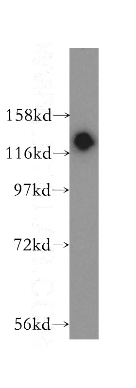
HeLa cells were subjected to SDS PAGE followed by western blot with Catalog No:113299(P130 antibody) at dilution of 1:500
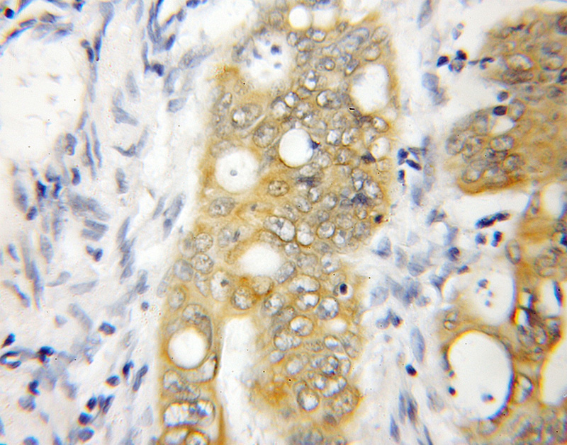
Immunohistochemical of paraffin-embedded human colon cancer using Catalog No:113299(P130 antibody) at dilution of 1:50 (under 10x lens)
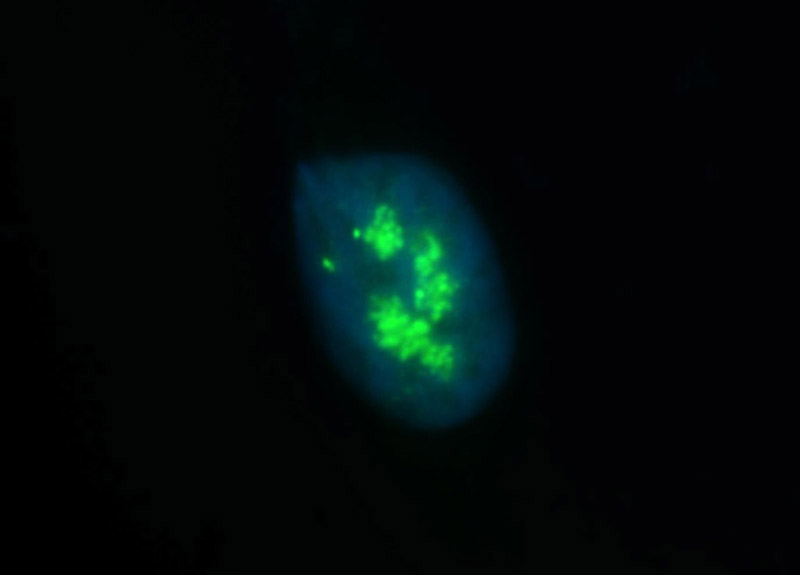
Immunofluorescent analysis of HepG2 cells, using NOLC1 antibody Catalog No:113299 at 1:50 dilution and FITC-labeled donkey anti-rabbit IgG(green). Blue pseudocolor = DAPI (fluorescent DNA dye).
IP Result of anti-P130 (IP:Catalog No:113299, 4ug; Detection:Catalog No:113299 1:500) with HeLa cells lysate 2000ug.
-
Background
Nucleolar and coiled-body phosphoprotein 1 (NOLC1) is a phosphoprotein that transiently associates with the mature nucleolar H/ACA and C/D box small nucleolar ribonucleoproteins (snoRNPs), guiding site-specific 2'-O-meth-ylation and pseudouridylation of pre-rRNAs [PMID:21266110]. It contains a nuclear localization signal binding sequence and is thought to shuttle between the nucleolus and the cytoplasm. NOLC1 plays an essential role in the synthesis of rRNA and the biosynthesis of ribosomes [PMID:8972203].
-
References
- Werner A, Iwasaki S, McGourty CA. Cell-fate determination by ubiquitin-dependent regulation of translation. Nature. 525(7570):523-7. 2015.
Related Products / Services
Please note: All products are "FOR RESEARCH USE ONLY AND ARE NOT INTENDED FOR DIAGNOSTIC OR THERAPEUTIC USE"