
-
Product Name
MT-ATP8 Polyclonal Antibody
- Documents
-
Description
Polyclonal antibody to MT-ATP8
-
Tested applications
WB, IHC, IF
-
Species reactivity
Human, Mouse, Rat
-
Alternative names
ATPase8 antibody; MTATP8 antibody; ATP8 antibody
-
Isotype
Rabbit IgG
-
Preparation
Antigen: Recombinant protein of human MT-ATP8.
-
Clonality
Polyclonal
-
Formulation
PBS with 0.02% sodium azide, 50% glycerol, pH7.3.
-
Storage instructions
Store at -20℃. Avoid freeze / thaw cycles.
-
Applications
WB 1:500 - 1:2000
IHC 1:50 - 1:100
IF 1:50 - 1:100 -
Validations
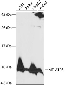
Western blot - MT-ATP8 Polyclonal Antibody
Western blot analysis of extracts of various cell lines, using MT-ATP8 antibody at 1:1000 dilution.Secondary antibody: HRP Goat Anti-Rabbit IgG (H+L) at 1:10000 dilution.Lysates/proteins: 25ug per lane.Blocking buffer: 3% nonfat dry milk in TBST.Detection: ECL Basic Kit .Exposure time: 2min.
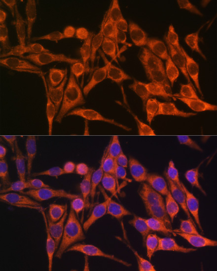
Immunofluorescence - MT-ATP8 Polyclonal Antibody
Immunofluorescence analysis of HeLa cells using MT-ATP8 antibody at dilution of 1:100. Blue: DAPI for nuclear staining.
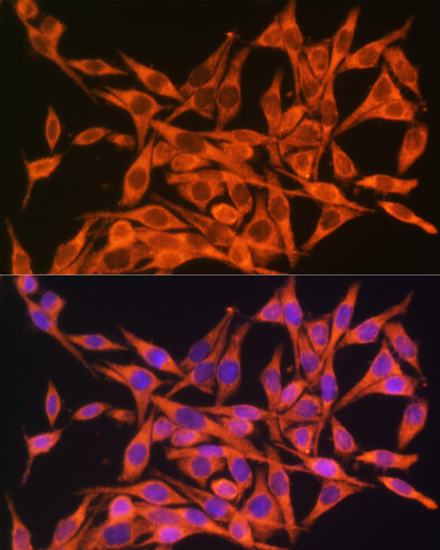
Immunofluorescence - MT-ATP8 Polyclonal Antibody
Immunofluorescence analysis of HeLa cells using MT-ATP8 antibody at dilution of 1:100. Blue: DAPI for nuclear staining.
-
Background
Mitochondrial membrane ATP synthase (F(1)F(0) ATP synthase or Complex V) produces ATP from ADP in the presence of a proton gradient across the membrane which is generated by electron transport complexes of the respiratory chain. F-type ATPases consist of two structural domains, F(1) - containing the extramembraneous catalytic core and F(0) - containing the membrane proton channel, linked together by a central stalk and a peripheral stalk. During catalysis, ATP synthesis in the catalytic domain of F(1) is coupled via a rotary mechanism of the central stalk subunits to proton translocation. Part of the complex F(0) domain. Minor subunit located with subunit a in the membrane (By similarity).
Related Products / Services
Please note: All products are "FOR RESEARCH USE ONLY AND ARE NOT INTENDED FOR DIAGNOSTIC OR THERAPEUTIC USE"