-
Product Name
IRAK1 Polyclonal Antibody
- Documents
-
Description
Polyclonal antibody to IRAK1
-
Tested applications
WB, IF
-
Species reactivity
Human, Mouse, Rat
-
Alternative names
IRAK1 antibody; IRAK antibody; pelle antibody; interleukin-1 receptor-associated kinase 1 antibody
-
Isotype
Rabbit IgG
-
Preparation
Antigen: Recombinant fusion protein containing a sequence corresponding to amino acids 424-633 of human IRAK1 (NP_001020414.1).
-
Clonality
Polyclonal
-
Formulation
PBS with 0.02% sodium azide, 50% glycerol, pH7.3.
-
Storage instructions
Store at -20℃. Avoid freeze / thaw cycles.
-
Applications
WB 1:1000 - 1:3000
IF 1:50 - 1:200 -
Validations

Western blot - IRAK1 Polyclonal Antibody
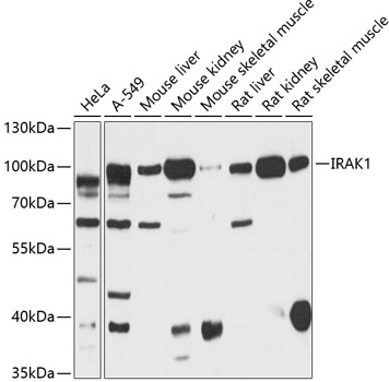
Western blot analysis of extracts of various cell lines, using IRAK1 antibody at 1:3000 dilution.Secondary antibody: HRP Goat Anti-Rabbit IgG (H+L) at 1:10000 dilution.Lysates/proteins: 25ug per lane.Blocking buffer: 3% nonfat dry milk in TBST.Detection: ECL Basic Kit .Exposure time: 5s.

Immunofluorescence - IRAK1 Polyclonal Antibody
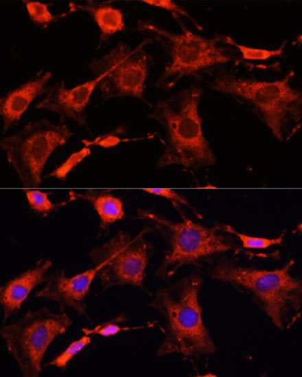
Immunofluorescence analysis of C6 cells using IRAK1 antibody at dilution of 1:100. Blue: DAPI for nuclear staining.

Immunofluorescence - IRAK1 Polyclonal Antibody
Immunofluorescence analysis of HeLa cells using IRAK1 antibody at dilution of 1:100. Blue: DAPI for nuclear staining.

Immunofluorescence - IRAK1 Polyclonal Antibody
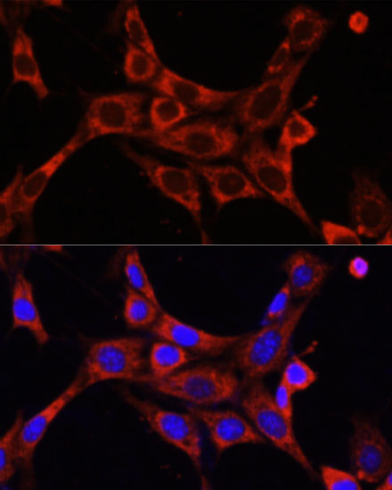
Immunofluorescence analysis of NIH/3T3 cells using IRAK1 antibody at dilution of 1:100. Blue: DAPI for nuclear staining.
-
Background
Serine/threonine-protein kinase that plays a critical role in initiating innate immune response against foreign pathogens. Involved in Toll-like receptor (TLR) and IL-1R signaling pathways. Is rapidly recruited by MYD88 to the receptor-signaling complex upon TLR activation. Association with MYD88 leads to IRAK1 phosphorylation by IRAK4 and subsequent autophosphorylation and kinase activation. Phosphorylates E3 ubiquitin ligases Pellino proteins (PELI1, PELI2 and PELI3) to promote pellino-mediated polyubiquitination of IRAK1. Then, the ubiquitin-binding domain of IKBKG/NEMO binds to polyubiquitinated IRAK1 bringing together the IRAK1-MAP3K7/TAK1-TRAF6 complex and the NEMO-IKKA-IKKB complex. In turn, MAP3K7/TAK1 activates IKKs (CHUK/IKKA and IKBKB/IKKB) leading to NF-kappa-B nuclear translocation and activation. Alternatively, phosphorylates TIRAP to promote its ubiquitination and subsequent degradation. Phosphorylates the interferon regulatory factor 7 (IRF7) to induce its activation and translocation to the nucleus, resulting in transcriptional activation of type I IFN genes, which drive the cell in an antiviral state. When sumoylated, translocates to the nucleus and phosphorylates STAT3.
Related Products / Services
Please note: All products are "FOR RESEARCH USE ONLY AND ARE NOT INTENDED FOR DIAGNOSTIC OR THERAPEUTIC USE"
