-
Product Name
EIF3D antibody
- Documents
-
Description
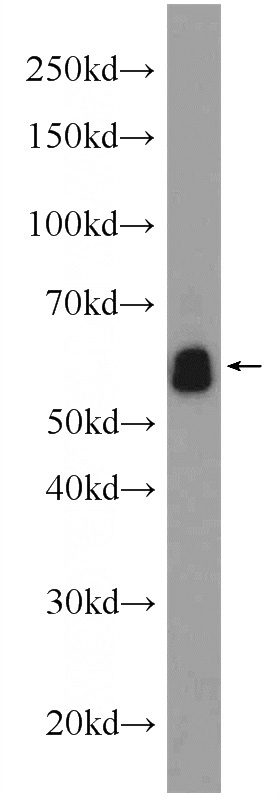
EIF3D Rabbit Polyclonal antibody. Positive IHC detected in human breast cancer tissue. Positive IF detected in MCF-7 cells, Ethacrynic acid treated HepG2 cells, HepG2 cells, transfected cells. Positive WB detected in mouse brain tissue, A549 cells, HepG2 cells, human brain tissue, L02 cells. Observed molecular weight by Western-blot: 66 kDa
-
Tested applications
ELISA, IF, IHC, WB
-
Species reactivity
Human, Mouse, Yeast; other species not tested.
-
Alternative names
eIF 3 zeta antibody; eIF3 p66 antibody; eIF3 zeta antibody; EIF3D antibody; EIF3S7 antibody
-
Isotype
Rabbit IgG
-
Preparation
This antibody was obtained by immunization of EIF3D recombinant protein (Accession Number: NM_003753). Purification method: Antigen affinity purified.
-
Clonality
Polyclonal
-
Formulation
PBS with 0.1% sodium azide and 50% glycerol pH 7.3.
-
Storage instructions
Store at -20℃. DO NOT ALIQUOT
-
Applications
Recommended Dilution:
WB: 1:200-1:2000
IHC: 1:20-1:200
IF: 1:20-1:200
-
Validations

mouse brain tissue were subjected to SDS PAGE followed by western blot with Catalog No:110192(EIF3D Antibody) at dilution of 1:600

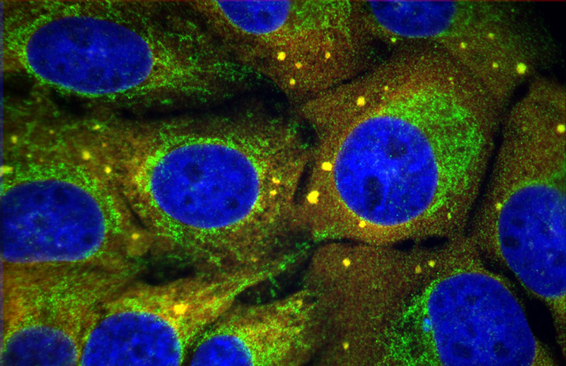
F result of Catalog No:110192(anti-EIF3D) in U2OS cell (treated with 100 mM sodium arsenite to cause stress-induced translational arrest) by Dr. Nancy Kedersha.U2OS cells (FAST-YFP stables; but not showing FAST-YFP); stained with PTG anti-eIF3p66 in green; and counterstained with anti-eIF3b (goat polyclonal) in red; nuclei stained blue using Hoechst.

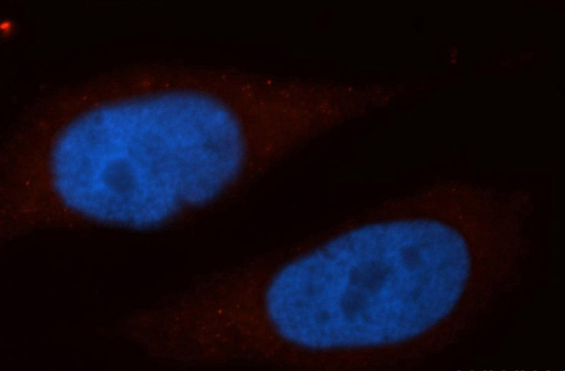
Immunofluorescent analysis of MCF-7 cells, using EIF3D antibody Catalog No:110192 at 1:50 dilution and Rhodamine-labeled goat anti-rabbit IgG (red). Blue pseudocolor = DAPI (fluorescent DNA dye).

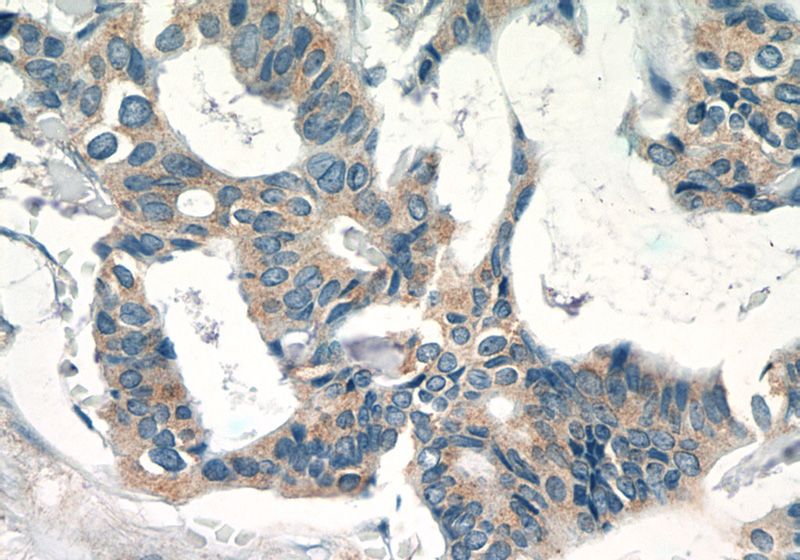
Immunohistochemistry of paraffin-embedded human breast cancer tissue slide using Catalog No:110192(EIF3D Antibody) at dilution of 1:50 (under 40x lens)
-
Background
The mammalian translation initiation factor 3 (eIF3), is a multiprotein complex of ~600 kDa that binds to the 40 S ribosome and promotes the binding of methionyl-tRNAi and mRNA. The EIF3S7(p66) is the major RNA binding subunit in this complex. Human eIF3-p66 shares 64% sequence identity with a hypothetical Caenorhabditis elegans protein, presumably the p66 homolog. Deletion analyses of recombinant derivatives of eIF3-p66 show that the RNA-binding domain lies within an N-terminal 71-amino acid region rich in lysine and arginine.
-
References
- Locker N, Easton LE, Lukavsky PJ. Affinity purification of eukaryotic 48S initiation complexes. RNA (New York, N.Y.). 12(4):683-90. 2006.
- Locker N, Lukavsky PJ. A practical approach to isolate 48S complexes: affinity purification and analyses. Methods in enzymology. 429:83-104. 2007.
- Locker N, Easton LE, Lukavsky PJ. HCV and CSFV IRES domain II mediate eIF2 release during 80S ribosome assembly. The EMBO journal. 26(3):795-805. 2007.
- Suo J, Snider SJ, Mills GB. Int6 regulates both proteasomal degradation and translation initiation and is critical for proper formation of acini by human mammary epithelium. Oncogene. 30(6):724-36. 2011.
- Otero JH, Suo J, Gordon C, Chang EC. Int6 and Moe1 interact with Cdc48 to regulate ERAD and proper chromosome segregation. Cell cycle (Georgetown, Tex.). 9(1):147-61. 2010.
- Rosario GX, Hondo E, Jeong JW. The LIF-mediated molecular signature regulating murine embryo implantation. Biology of reproduction. 91(3):66. 2014.
- Leen EN, Sorgeloos F, Correia S. A Conserved Interaction between a C-Terminal Motif in Norovirus VPg and the HEAT-1 Domain of eIF4G Is Essential for Translation Initiation. PLoS pathogens. 12(1):e1005379. 2016.
Related Products / Services
Please note: All products are "FOR RESEARCH USE ONLY AND ARE NOT INTENDED FOR DIAGNOSTIC OR THERAPEUTIC USE"
