-
Product Name
CFI Polyclonal Antibody
- Documents
-
Description
Polyclonal antibody to CFI
-
Tested applications
WB, IF
-
Species reactivity
Human, Mouse
-
Alternative names
CFI antibody; AHUS3 antibody; ARMD13 antibody; C3BINA antibody; C3b-INA antibody; FI antibody; IF antibody; KAF antibody; complement factor I antibody
-
Isotype
Rabbit IgG
-
Preparation
Antigen: Recombinant fusion protein containing a sequence corresponding to amino acids 19-300 of human CFI (NP_000195.2).
-
Clonality
Polyclonal
-
Formulation
PBS with 0.02% sodium azide, 50% glycerol, pH7.3.
-
Storage instructions
Store at -20℃. Avoid freeze / thaw cycles.
-
Applications
WB 1:500 - 1:2000
IF 1:50 - 1:100 -
Validations

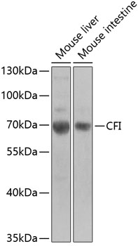
Western blot - CFI Polyclonal Antibody
Western blot analysis of extracts of various cell lines, using CFI antibody at 1:1000 dilution.Secondary antibody: HRP Goat Anti-Rabbit IgG (H+L) at 1:10000 dilution.Lysates/proteins: 25ug per lane.Blocking buffer: 3% nonfat dry milk in TBST.Detection: ECL Basic Kit .Exposure time: 90s.

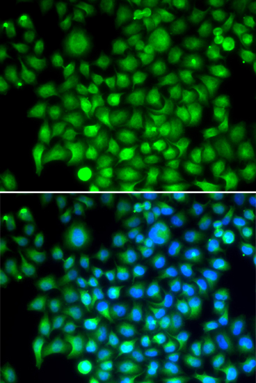
Immunofluorescence - CFI Polyclonal Antibody
Immunofluorescence analysis of HeLa cells using CFI antibody . Blue: DAPI for nuclear staining.
-
Background
Trypsin-like serine protease that plays an essential role in regulating the immune response by controlling all complement pathways. Inhibits these pathways by cleaving three peptide bonds in the alpha-chain of C3b and two bonds in the alpha-chain of C4b thereby inactivating these proteins. Essential cofactors for these reactions include factor H and C4BP in the fluid phase and membrane cofactor protein/CD46 and CR1 on cell surfaces. The presence of these cofactors on healthy cells allows degradation of deposited C3b by CFI in order to prevent undesired complement activation, while in apoptotic cells or microbes, the absence of such cofactors leads to C3b-mediated complement activation and subsequent opsonization.
Related Products / Services
Please note: All products are "FOR RESEARCH USE ONLY AND ARE NOT INTENDED FOR DIAGNOSTIC OR THERAPEUTIC USE"
