-
Product Name
CDC6 antibody
- Documents
-
Description
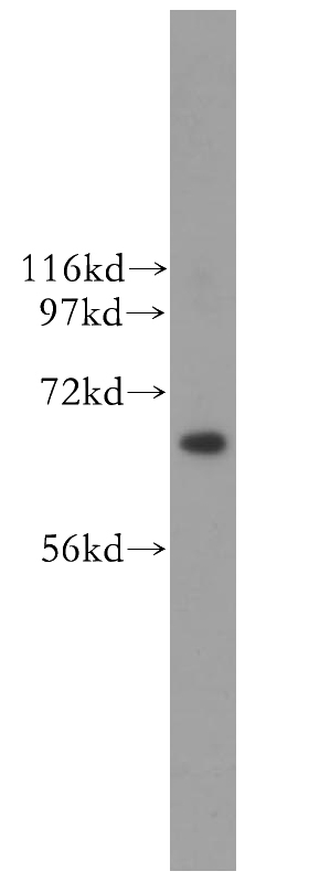
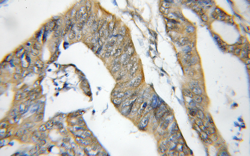
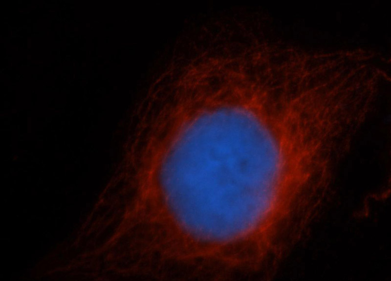
CDC6 Rabbit Polyclonal antibody. Positive WB detected in HeLa cells, Jurkat cells, SMMC-7721 cells. Positive IHC detected in human colon cancer tissue, human colon tissue. Positive IF detected in HepG2 cells. Observed molecular weight by Western-blot: 63 kDa
-
Tested applications
ELISA, WB, IHC, IF
-
Species reactivity
Human,Mouse,Rat; other species not tested.
-
Alternative names
Cdc18 related protein antibody; CDC18L antibody; CDC6 antibody; CDC6 related protein antibody; HsCDC18 antibody; HsCDC6 antibody; p62(cdc6) antibody
-
Isotype
Rabbit IgG
-
Preparation
This antibody was obtained by immunization of CDC6 recombinant protein (Accession Number: NM_001254). Purification method: Antigen affinity purified.
-
Clonality
Polyclonal
-
Formulation
PBS with 0.1% sodium azide and 50% glycerol pH 7.3.
-
Storage instructions
Store at -20℃. DO NOT ALIQUOT
-
Applications
Recommended Dilution:
WB: 1:1000-1:10000
IHC: 1:20-1:200
IF: 1:20-1:200
-
Validations

HeLa cells were subjected to SDS PAGE followed by western blot with Catalog No:109111(CDC6 antibody) at dilution of 1:400

Immunohistochemical of paraffin-embedded human colon cancer using Catalog No:109111(CDC6 antibody) at dilution of 1:50 (under 10x lens)

Immunofluorescent analysis of HepG2 cells, using CDC6 antibody Catalog No:109111 at 1:50 and Rhodamine-labeled goat anti-rabbit IgG (red). Blue pseudocolor = DAPI (fluorescent DNA dye).
-
Background
CDC6, also named as CDC18L and HsCDC18, belongs to the CDC6/cdc18 family. It is involved in the initiation of DNA replication and functions as a regulator at the early steps of DNA replication. The active Cdc6 functioning as a dimer at replication origins (10793143,15096526). It also participates in checkpoint controls that ensure DNA replication is completed before mitosis is initiated. It localizes in cell nucleus during cell cyle G1, but translocates to the cytoplasm at the start of S phase. The subcellular translocation of this protein during cell cyle is regulated through its phosphorylation by Cdks. Defects in CDC6 are the cause of Meier-Gorlin syndrome type 5 (MGORS5). MGORS5 is a syndrome characterized by bilateral microtia, aplasia/hypoplasia of the patellae, and severe intrauterine and postnatal growth retardation with short stature and poor weight gain. This antibody is a rabbit polyclonal antibody raised against residues near the C terminus of human CDC6.
-
References
- Basaki Y, Taguchi K, Izumi H. Y-box binding protein-1 (YB-1) promotes cell cycle progression through CDC6-dependent pathway in human cancer cells. European journal of cancer (Oxford, England : 1990). 46(5):954-65. 2010.
- Yim H, Erikson RL. Cell division cycle 6, a mitotic substrate of polo-like kinase 1, regulates chromosomal segregation mediated by cyclin-dependent kinase 1 and separase. Proceedings of the National Academy of Sciences of the United States of America. 107(46):19742-7. 2010.
- Akiba J, Murakami Y, Noda M. N-myc downstream regulated gene1/Cap43 overexpression suppresses tumor growth by hepatic cancer cells through cell cycle arrest at the G0/G1 phase. Cancer letters. 310(1):25-34. 2011.
- Ma P, Schultz RM. Histone deacetylase 2 (HDAC2) regulates chromosome segregation and kinetochore function via H4K16 deacetylation during oocyte maturation in mouse. PLoS genetics. 9(3):e1003377. 2013.
- Harada M, Kotake Y, Ohhata T. YB-1 promotes transcription of cyclin D1 in human non-small-cell lung cancers. Genes to cells : devoted to molecular & cellular mechanisms. 19(6):504-16. 2014.
Related Products / Services
Please note: All products are "FOR RESEARCH USE ONLY AND ARE NOT INTENDED FOR DIAGNOSTIC OR THERAPEUTIC USE"
