
-
Product Name
CD3 delta antibody
- Documents
-
Description
CD3 delta Rabbit Polyclonal antibody. Positive FC detected in Jurkat cells. Positive IHC detected in human tonsillitis tissue, human lymphoma tissue. Positive IP detected in Jurkat cells. Positive WB detected in Jurkat cells, mouse thymus tissue. Observed molecular weight by Western-blot: 20-25 kDa
-
Tested applications
ELISA, WB, FC, IHC, IP
-
Species reactivity
Human, Mouse; other species not tested.
-
Alternative names
CD3 delta antibody; CD3D antibody; CD3-delta antibody; T cell receptor T3 delta chain antibody; T3D antibody
-
Isotype
Rabbit IgG
-
Preparation
This antibody was obtained by immunization of CD3 delta recombinant protein (Accession Number: XM_017018543). Purification method: Antigen affinity purified.
-
Clonality
Polyclonal
-
Formulation
PBS with 0.02% sodium azide and 50% glycerol pH 7.3.
-
Storage instructions
Store at -20℃. DO NOT ALIQUOT
-
Applications
Recommended Dilution:
WB: 1:200-1:2000
IP: 1:200-1:2000
IHC: 1:20-1:200
-
Validations
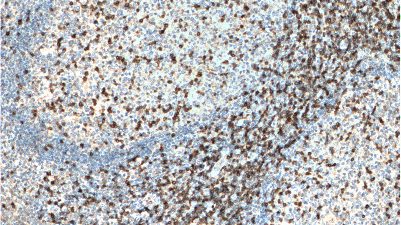
Immunohistochemistry of paraffin-embedded human tonsillitis tissue slide using Catalog No:109019(CD3D Antibody) at dilution of 1:200 (under 10x lens). heat mediated antigen retrieved with Tris-EDTA buffer(pH9).
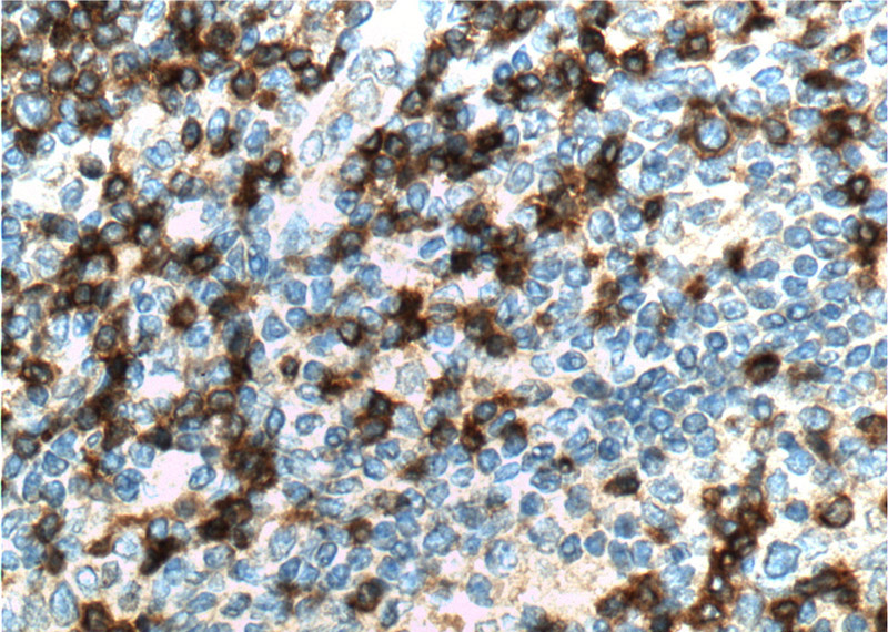
Immunohistochemistry of paraffin-embedded human tonsillitis tissue slide using Catalog No:109019(CD3D Antibody) at dilution of 1:200 (under 40x lens). heat mediated antigen retrieved with Tris-EDTA buffer(pH9).
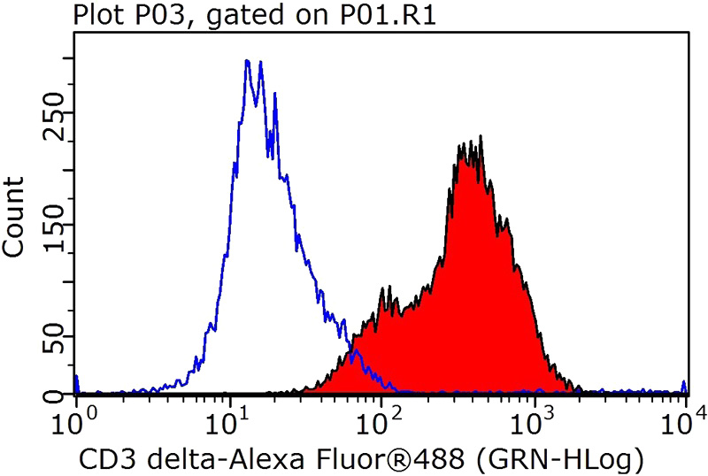
1X10^6 Jurkat cells were stained with 0.5ug CD3D antibody (Catalog No:109019, red) and control antibody (blue). Fixed with 90% MeOH blocked with 3% BSA (30 min). Alexa Fluor 488-congugated AffiniPure Goat Anti-Rabbit IgG(H+L) with dilution 1:1000.
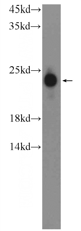
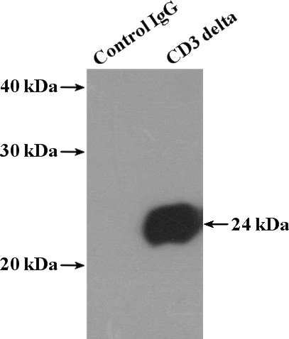
Jurkat cells were subjected to SDS PAGE followed by western blot with Catalog No:109019(CD3D antibody) at dilution of 1:800
IP Result of anti-CD3D (IP:Catalog No:109019, 4ug; Detection:Catalog No:109019 1:800) with Jurkat cells lysate 2800ug.
-
Background
CD3 is a complex of proteins that directly associates with the T cell receptor (TCR). The TCR/CD3 complex of T-lymphocytes consists of either a TCR alpha/beta or TCR gamma/delta heterodimer coexpressed at the cell surface with the invariant subunits of CD3 labeled gamma, delta, epsilon, zeta, and eta. The TCR recognizes antigens bound to major histocompatibility complex (MHC) molecules. TCR-mediated peptide-MHC recognition is transmitted to the CD3 complex, leading to the intracellular signal transduction. CD3 is considered to be a pan-T cell marker for detection of normal and neoplastic T cells.
Related Products / Services
Please note: All products are "FOR RESEARCH USE ONLY AND ARE NOT INTENDED FOR DIAGNOSTIC OR THERAPEUTIC USE"