

-
Product Name
Caspase 9/p35/p10 antibody
- Documents
-
Description
Caspase 9/p35/p10 Rabbit Polyclonal antibody. Positive FC detected in HepG2 cells. Positive IHC detected in mouse lung tissue, human heart tissue. Positive IF detected in HepG2 cells. Positive WB detected in HeLa cells, HL-60 cells, Jurkat cells. Positive IP detected in HeLa cells. Observed molecular weight by Western-blot: 46 kDa
-
Tested applications
ELISA, IF, IHC, IP, WB, FC
-
Species reactivity
Human,Mouse,Rat; other species not tested.
-
Alternative names
APAF 3 antibody; APAF3 antibody; Apoptotic protease Mch 6 antibody; CASP 9 antibody; CASP9 antibody; Caspase 9 antibody; CASPASE 9c antibody; Caspase9 antibody; Caspase-9 antibody; ICE LAP6 antibody; ICE like apoptotic protease 6 antibody; MCH6 antibody
-
Isotype
Rabbit IgG
-
Preparation
This antibody was obtained by immunization of Caspase 9/p35/p10 recombinant protein (Accession Number: NM_001229). Purification method: Antigen affinity purified.
-
Clonality
Polyclonal
-
Formulation
PBS with 0.1% sodium azide and 50% glycerol pH 7.3.
-
Storage instructions
Store at -20℃. DO NOT ALIQUOT
-
Applications
Recommended Dilution:
WB: 1:500-1:5000
IP: 1:200-1:1000
IHC: 1:10-1:100
IF: 1:10-1:100
-
Validations
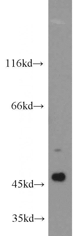
HeLa cells were subjected to SDS PAGE followed by western blot with Catalog No:108881(CASP9 antibody) at dilution of 1:1000
IP Result of anti-CASP9 (IP:Catalog No:108881, 3ug; Detection:Catalog No:108881 1:200) with HeLa cells lysate 2500ug.
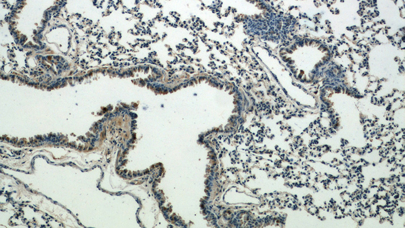
Immunohistochemical of paraffin-embedded mouse lung using Catalog No:108881(CASP9 antibody) at dilution of 1:25 (under 10x lens)
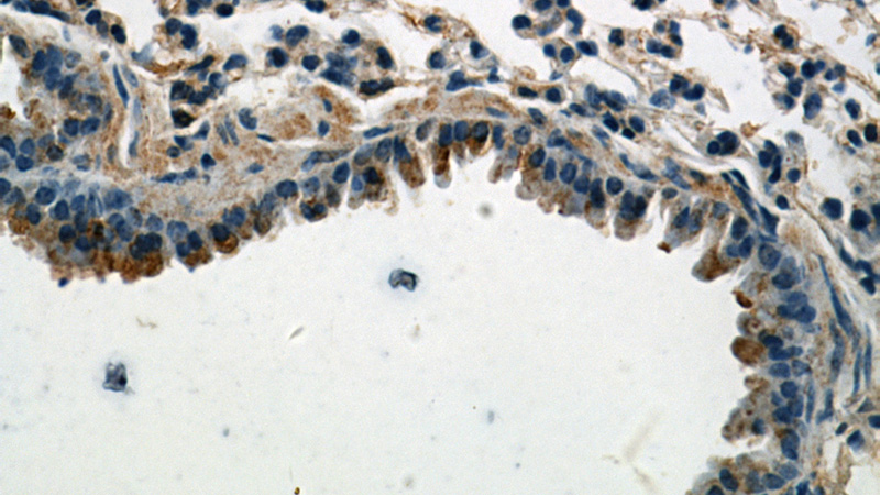
Immunohistochemical of paraffin-embedded mouse lung using Catalog No:108881(CASP9 antibody) at dilution of 1:25 (under 40x lens)
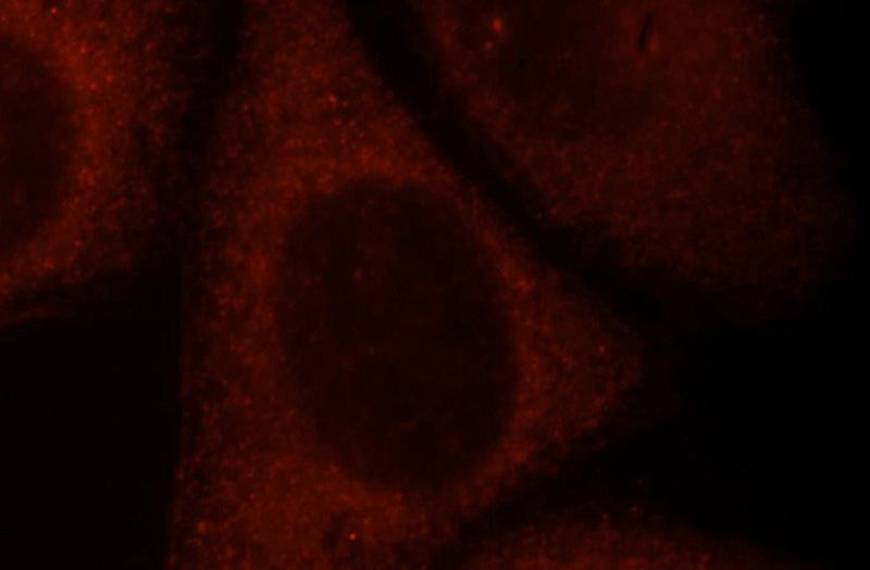
Immunofluorescent analysis of HepG2 cells, using CASP9 antibody Catalog No:108881 at 1:25 dilution and Rhodamine-labeled goat anti-rabbit IgG (red).
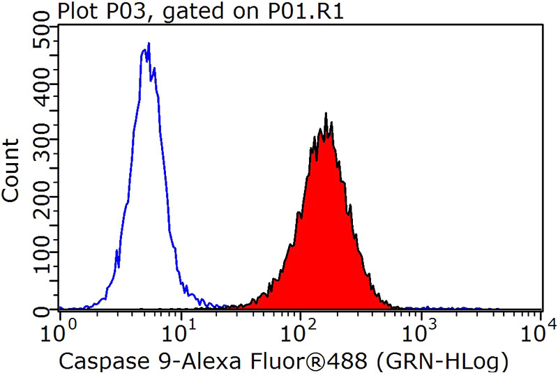
1X10^6 HepG2 cells were stained with 0.2ug Caspase 9 antibody (Catalog No:108881, red) and control antibody (blue). Fixed with 90% MeOH blocked with 3% BSA (30 min). Alexa Fluor 488-congugated AffiniPure Goat Anti-Rabbit IgG(H+L) with dilution 1:1000.
-
Background
Caspase 9, apoptosis-related cysteine protease (CASP9, synonyms: MCH6, APAF3, APAF-3, ICE-LAP6, CASPASE-9c)is a member of the cysteine-aspartic acid protease (caspase) family. Sequential activation of caspases plays a central role in the execution-phase of cell apoptosis. Caspases exist as inactive proenzymes which undergo proteolytic processing at conserved aspartic residues to produce 2 subunits, large and small, that dimerize to form the active enzyme. Caspase 9 is processed by APAF1; this step is thought to be one of the earliest in the caspase activation cascade.
-
References
- Chen W, Hou J, Yin Y. alpha-Bisabolol induces dose- and time-dependent apoptosis in HepG2 cells via a Fas- and mitochondrial-related pathway, involves p53 and NFkappaB. Biochemical pharmacology. 80(2):247-54. 2010.
- Liu H, Xiao Y, Xiong C, Wei A, Ruan J. Apoptosis induced by a new flavonoid in human hepatoma HepG2 cells involves reactive oxygen species-mediated mitochondrial dysfunction and MAPK activation. European journal of pharmacology. 654(3):209-16. 2011.
- Zheng S, Chang S, Lu J. Characterization of 9-nitrocamptothecin liposomes: anticancer properties and mechanisms on hepatocellular carcinoma in vitro and in vivo. PloS one. 6(6):e21064. 2011.
- Yin Y, Chen W, Tang C. NF-κB, JNK and p53 pathways are involved in tubeimoside-1-induced apoptosis in HepG2 cells with oxidative stress and G₂/M cell cycle arrest. Food and chemical toxicology : an international journal published for the British Industrial Biological Research Association. 49(12):3046-54. 2011.
- Lei JC, Yu JQ, Yin Y, Liu YW, Zou GL. Alantolactone induces activation of apoptosis in human hepatoma cells. Food and chemical toxicology : an international journal published for the British Industrial Biological Research Association. 50(9):3313-9. 2012.
- Yu JQ, Bao W, Lei JC. Emodin regulates apoptotic pathway in human liver cancer cells. Phytotherapy research : PTR. 27(2):251-7. 2013.
- Jia Y, Dong X, Zhou P. The synthesis and biological evaluation of novel Danshensu-cysteine analog conjugates as cardiovascular-protective agents. European journal of medicinal chemistry. 55:176-87. 2012.
- Yin X, Yu B, Tang Z. Bifidobacterium infantis-mediated HSV-TK/GCV suicide gene therapy induces both extrinsic and intrinsic apoptosis in a rat model of bladder cancer. Cancer gene therapy. 20(2):77-81. 2013.
Related Products / Services
Please note: All products are "FOR RESEARCH USE ONLY AND ARE NOT INTENDED FOR DIAGNOSTIC OR THERAPEUTIC USE"