
-
Product Name
beta Actin antibody
- Documents
-
Description
beta Actin Rabbit Polyclonal antibody. Positive WB detected in HeLa cells, A431 cells, A549 cells, C6 cells, HEK-293 cells, HepG2 cells, human testis tissue, Jurkat cells, MCF7 cells, mouse brain tissue, mouse lung tissue, mouse spleen tissue, mouse uterus tissue, NIH/3T3 cells, rat brain tissue, SGC-7901 cells, Sp2/0 cells. Positive IF detected in HepG2 cells. Positive IHC detected in human skeletal muscle tissue, human colon tissue, human heart tissue, human kidney tissue. Observed molecular weight by Western-blot: 42 kDa
-
Tested applications
ELISA, IF, WB, IHC
-
Species reactivity
Human,Mouse,Rat,Zebrafish, Monkey; other species not tested.
-
Alternative names
ACTB antibody; Actin antibody; actin antibody; beta antibody; Actin antibody; cytoplasmic 1 antibody; B actin antibody; Beta actin antibody; F actin antibody; PS1TP5BP1 antibody; β actin antibody
-
Isotype
Rabbit IgG
-
Preparation
This antibody was obtained by immunization of beta Actin recombinant protein (Accession Number: NM_001101). Purification method: Antigen affinity purified.
-
Clonality
Polyclonal
-
Formulation
PBS with 0.02% sodium azide and 50% glycerol pH 7.3.
-
Storage instructions
Store at -20℃. DO NOT ALIQUOT
-
Applications
Recommended Dilution:
WB: 1:1000-1:10000
IHC: 1:20-1:200
IF: 1:10-1:100
-
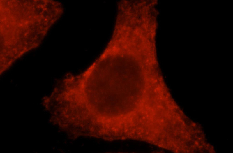
Validations
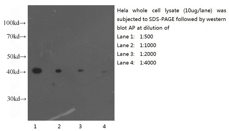
Western blot of Hela cell with anti-Actin-Beta (Catalog No:117305) at various dilutions.
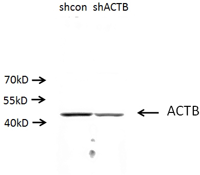
A549 cells (shcontrol and shRNA of Beta actin) were subjected to SDS PAGE followed by western blot with Catalog No:117305(ACTB antibody) at dilution of 1:500. (Data provided by Angran Biotech (www.miRNAlab.com)).
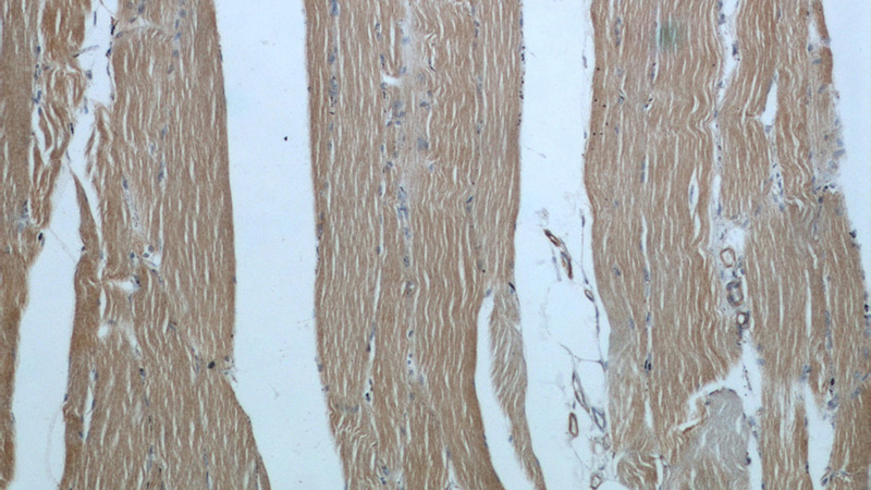
Immunohistochemical of paraffin-embedded human skeletal muscle using Catalog No:117305(ACTB antibody) at dilution of 1:100 (under 10x lens)
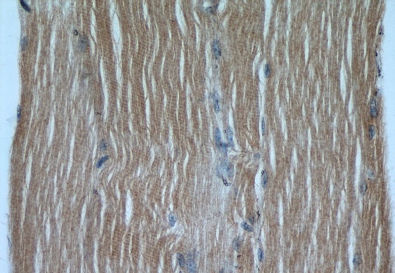
Immunohistochemical of paraffin-embedded human skeletal muscle using Catalog No:117305(ACTB antibody) at dilution of 1:100 (under 40x lens)
Immunofluorescent analysis of HepG2 cells, using ACTB antibody Catalog No:117305 at 1:25 dilution and Rhodamine-labeled goat anti-rabbit IgG (red).
-
Background
Beta actin, also named as ACTB and F-Actin, belongs to the actin family. Actins are highly conserved globular proteins that are involved in various types of cell motility and are ubiquitously expressed in all eukaryotic cells. At least six isoforms of actins are known in mammals and other vertebrates: alpha (ACTC1, cardiac muscle 1), alpha 1 (ACTA1, skeletal muscle) and 2 (ACTA2, aortic smooth muscle), beta (ACTB), gamma 1 (ACTG1) and 2 (ACTG2, enteric smooth muscle). Beta and gamma 1 are two non-muscle actin proteins. Most actins consist of 376aa, while ACTG2 (rich in muscles) has 375aa and ACTG1(found in non-muscle cells) has only 374aa. Beta actin has been widely used as the internal control in RT-PCR and Western Blotting as a 42-kDa protein. However, the 41 kDa cleaved fragment of beta actin can be generated during apoptosis process. This antibody was generated against N-terminal region of human beta actin protein and can cross-react with other actins.
-
References
- Xu C, Wang P, Liu Y. Integrative genomics in combination with RNA interference identifies prognostic and functionally relevant gene targets for oral squamous cell carcinoma. PLoS genetics. 9(1):e1003169. 2013.
- Weber H, Müller L, Jonas L, Schult C, Sparmann G, Schuff-Werner P. Calpain mediates caspase-dependent apoptosis initiated by hydrogen peroxide in pancreatic acinar AR42J cells. Free radical research. 47(5):432-46. 2013.
- Melià MJ, Kubota A, Ortolano S. Limb-girdle muscular dystrophy 1F is caused by a microdeletion in the transportin 3 gene. Brain : a journal of neurology. 136(Pt 5):1508-17. 2013.
- Zhuo L, Liu Q, Liu L. Roles of 3,4-methylenedioxymethamphetamine (MDMA)-induced alteration of connexin43 and intracellular Ca(2+) oscillation in its cardiotoxicity. Toxicology. 310:61-72. 2013.
- Luo J, Zhou H, Wang F. The hepatitis B virus X protein downregulates NF-κB signaling pathways through decreasing the Notch signaling pathway in HBx-transformed L02 cells. International journal of oncology. 42(5):1636-43. 2013.
- Xu K, Gao J, Yang X, Yao Y, Liu Q. Cytohesin-2 as a novel prognostic marker for hepatocellular carcinoma. Oncology reports. 29(6):2211-8. 2013.
- Xie HX, Lu JF, Rolhion N. Edwardsiella tarda-Induced cytotoxicity depends on its type III secretion system and flagellin. Infection and immunity. 82(8):3436-45. 2014.
- Shi J, Duan Z, Sun J. Identification and validation of a novel microRNA-like molecule derived from a cytoplasmic RNA virus antigenome by bioinformatics and experimental approaches. Virology journal. 11:121. 2014.
Related Products / Services
Please note: All products are "FOR RESEARCH USE ONLY AND ARE NOT INTENDED FOR DIAGNOSTIC OR THERAPEUTIC USE"